. 2022 Jun 17;6(1):26.
doi: 10.1186/s41747-022-00283-z.

Twinkling-guided ultrasound detection of polymethyl methacrylate as a potential breast biopsy marker: a comparative investigation

Affiliations

Twinkling-guided ultrasound detection of polymethyl methacrylate as a potential breast biopsy marker: a comparative investigation

Christine U Lee et al. Eur Radiol Exp. .

Abstract

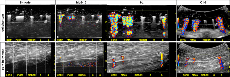
Since its first description 25 years ago, color Doppler twinkling has been a compelling ultrasound feature in diagnosing urinary stones. While the fundamental cause of twinkling remains elusive, the distinctive twinkling signature is diagnostically valuable in clinical practice. It can be inferred that if an entity twinkles, it empirically has certain physical features. This work investigates a manipulable polymeric material, polymethyl methacrylate (PMMA), which twinkles and has measurable surface roughness and porosity that likely contribute to twinkling. Comparative investigation of these structural properties and of the twinkling signatures of breast biopsy markers made from PMMA and selected commercially available markers showed how twinkling can improve ultrasound detection of devices intentionally designed to twinkle. While this specific application of detecting breast biopsy markers by twinkling may provide a way to approach an unmet need in the care of patients with breast cancer, this work ultimately provides a platform from which the keys to unlocking the fundamental physics of twinkling can be rigorously explored.

Keywords: Artifact; Polymethyl methacrylate; Porosity; Surface properties; Ultrasonography.


Conflict of interest statement

The authors declare that they have no competing interests.

Figures

Fig. 1
Fig. 1
Ultrasound of five markers (cork, PMMA, ribbon, O, and Q, from left to right) are seen on B-mode imaging in a gel phantom (a) and in ex vivo pork belly meat (e). For the cork, PMMA, and Q markers, the ML6-15 transducer demonstrated the weakest twinkling signatures, particularly evident in the pork belly study (b, f, Supplemental Materials B and F). While the 9L transducer showed exuberant, persistent twinkling signatures for the cork, PMMA, and Q markers in the gel phantom (c, Supplemental Materials C1 and C2), the twinkling was present but reduced in the pork belly meat (g, Supplemental Materials G1 and G2). The C1-6 transducer shows persistent, exuberant twinkling for the cork, PMMA, and Q markers in the gel phantom (d, Supplemental Material D) and pork belly meat (h, Supplemental Material H). The ribbon and O markers had a twinkling score of 0. The five markers were not perfectly aligned along a line so could not be optimally depicted in a single image; the white dotted vertical lines (c, e, f, g) indicate spliced frames from the cine clip providing optimal marker visualization. The thin echogenic parallel line in the gel phantom (a, b, c, d) is the interface of two stacked gel phantoms to minimize back scatter from the tabletop. PMMA Polymethyl methacrylate
Fig. 2
Fig. 2
Marker characteristics on ultrasound, SEM, optical profile, and micro-CT. Color Doppler twinkling signatures of markers (1st column), and their surface features from SEM at 50 × magnification (2nd column) correlate with what was predicted by surface roughness measurements (3rd column). Additionally, micro-computed tomography (4th column) provided a metric for porosity for the TriMark® cork and PMMA markers (5th column). Based on areal surface roughness measurements (see Fig. 3), the PMMA marker could be predicted to twinkle on color Doppler ultrasound. Both the PMMA marker and the TriMark® cork were rated as 4+ twinkling, surpassing expectations. A frequently used marker, the UltraClip™ ribbon clip, was rated 0 twinkling and did not have appreciable surface roughness. PMMA Polymethyl methacrylate, SEM Scanning electron microscopy
Fig. 3
Fig. 3
Surface roughness versus twinkling. Boxplots of areal surface roughness (Sa) measurements (y-axis, log-scale) with individual observations plotted, grouped by consensus absence (twinkling score 0) or presence of twinkling (score ≥ 3). Boxplots are slightly jittered horizontally solely to avoid direct overlap. Coloration and plotting symbols are separately designated for each marker per the legend. Figure produced using R v3.6.1 (R Foundation). Cork TriMark®, O SenoMark™, PMMA Polymethyl methacrylate, Q Tumark®, Ribbon UltraClip™

References

    1. Rahmouni A, Bargoin R, Herment A, Bargoin N, Vasile N. Color Doppler twinkling artifact in hyperechoic regions. Radiology. 1996;199:269–271. doi: 10.1148/radiology.199.1.8633158. - DOI - PubMed
    1. Lee JY, Kim SH, Cho JY, Han D. Color and power Doppler twinkling artifacts from urinary stones: clinical observations and phantom studies. AJR Am J Roentgenol. 2001;176:1441–1445. doi: 10.2214/ajr.176.6.1761441. - DOI - PubMed
    1. Shang M, Sun X, Liu Q, et al. Quantitative evaluation of the effects of urinary stone composition and size on color Doppler twinkling artifact: a phantom study. J Ultrasound Med. 2017;36:733–740. doi: 10.7863/ultra.16.01039. - DOI - PubMed
    1. Simon JC, Sapozhnikov OA, Kreider W, Breshock M, Williams JC, Bailey MR. The role of trapped bubbles in kidney stone detection with the color Doppler ultrasound twinkling artifact. Phys Med Biol. 2018;63:025011. doi: 10.1088/1361-6560/aa9a2f. - DOI - PMC - PubMed
    1. Wang M, Li J, Xiao J, Shi D, Zhang K. Systematic analysis of factors related to display of the twinkling artifact by a phantom: an optimized investigation. J Ultrasound Med. 2011;30:1449–1457. doi: 10.7863/jum.2011.30.11.1449. - DOI - PubMed

Publication types

Substances

LinkOut - more resources