

Reliability and Validity of Standing Lateral Radiograph Method for Measuring Acetabular Component Version: A Modified Cross-table Lateral Radiograph Method
- PMID: 35711111
- PMCID: PMC9363715
- DOI: 10.1111/os.13373
Reliability and Validity of Standing Lateral Radiograph Method for Measuring Acetabular Component Version: A Modified Cross-table Lateral Radiograph Method
Abstract
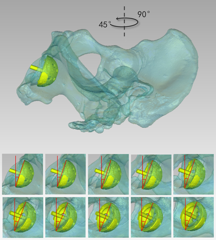
Objectives: To investigate the effect of the X-ray incidence angle on cup version measurements and the reliability and validity of standing lateral (SL) radiography for measuring cup versions.
Methods: Cup versions under different X-ray incidence angles were investigated by the 3D simulation analysis. Ninety-three patients, who underwent primary total hip arthroplasty (THA) with postoperative SL radiographs and CT scans between April 2020 and December 2021, were retrospectively analyzed. SL radiography was taken under naturally standing position, correcting for the measurement error of pelvic tilt in cross-table lateral (CL) radiography. Cup versions were measured on SL radiographs and CT images by two qualified orthopedic physicians. The intra- and inter-observer reliabilities were assessed by intra-class correlation coefficient. The consistency between radiographic and CT measurements was evaluated using Pearson correlation coefficient.
Results: No significant differences in cup version measurements were observed between groups of different X-ray incidence angles (P = 0.663) in the 3D simulation analysis. All measurements had excellent intra- and inter-observer reliabilities, with an intraclass correlation coefficient of >0.95. Mean cup version measurements from SL radiographs correlated well with those from CT scans (r = 0.853, P < 0.001). The mean difference between radiographic and CT measurements was -0.49° (range -12.62° to 10.37°, SD 3.95°), and the majority of differences were within the 95% limits of agreement.
Conclusion: The cup versions measured with SL radiography were close to the CT measurements. SL radiograph method is reliable and valid for measuring acetabular component version after THA.
Keywords: Acetabular component version; Cross-table lateral radiograph; Hip dislocation; Standing lateral radiograph; Total hip arthroplasty.
© 2022 The Authors. Orthopaedic Surgery published by Tianjin Hospital and John Wiley & Sons Australia, Ltd.
Figures


References
-
- Hevesi M, Wyles CC, Rouzrokh P, Erickson BJ, Maradit‐Kremers H, Lewallen DG, et al. Redefining the 3D topography of the acetabular safe zone: a multivariable study evaluating prosthetic hip stability. J Bone Joint Surg Am. 2022;104:239–45. - PubMed
-
- Grammatopoulos G, Thomas GE, Pandit H, Beard DJ, Gill HS, Murray DW. The effect of orientation of the acetabular component on outcome following total hip arthroplasty with small diameter hard‐on‐soft bearings. Bone Joint J. 2015;97:164–72. - PubMed
-
- McKnight BM, Trasolini NA, Dorr LD. Spinopelvic motion and impingement in total hip arthroplasty. J Arthroplasty. 2019;34:S53–s6. - PubMed
-
- Phillips CB, Barrett JA, Losina E, Mahomed NN, Lingard EA, Guadagnoli E, et al. Incidence rates of dislocation, pulmonary embolism, and deep infection during the first six months after elective total hip replacement. J Bone Joint Surg Am. 2003;85:20–6. - PubMed
MeSH terms
Grants and funding
- 2019M663265/National Natural Science Foundation of China
- 2019TQ0387/National Natural Science Foundation of China
- 2019M663265/China Postdoctoral Science Foundation funded project
- 2019TQ0387/China Postdoctoral Science Foundation funded project
- 2019A1515011647/Guangdong Basic and Applied Basic Research Foundation
LinkOut - more resources
Full Text Sources
Medical
Miscellaneous
