

Mechanistic Insights Into Inflammation-Induced Arrhythmias: A Simulation Study
- PMID: 35711306
- PMCID: PMC9196871
- DOI: 10.3389/fphys.2022.843292
Mechanistic Insights Into Inflammation-Induced Arrhythmias: A Simulation Study
Abstract
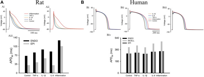
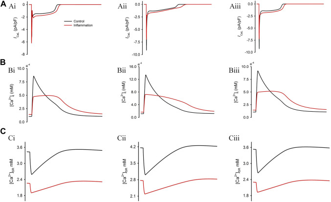
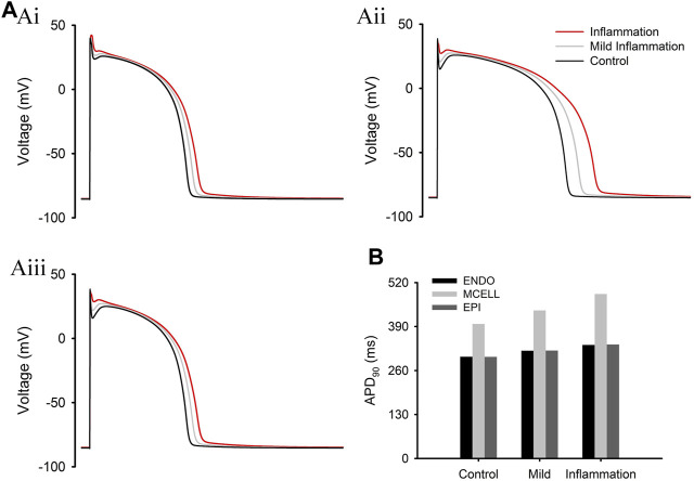
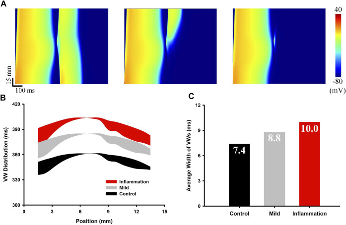
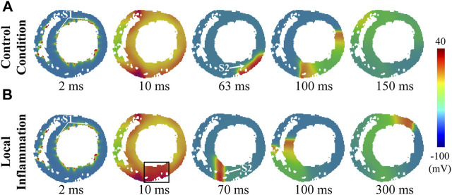
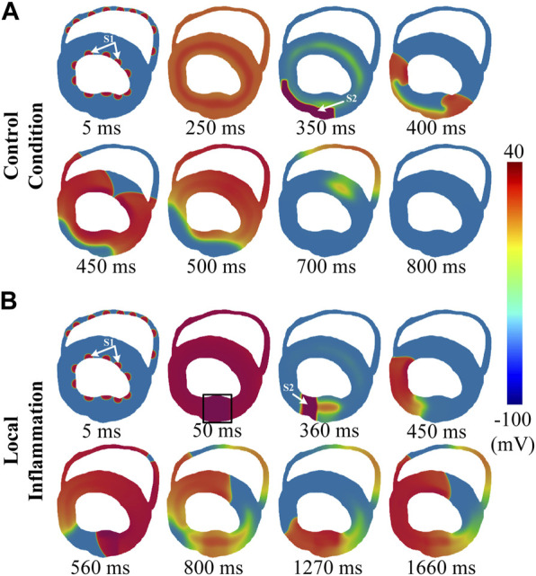
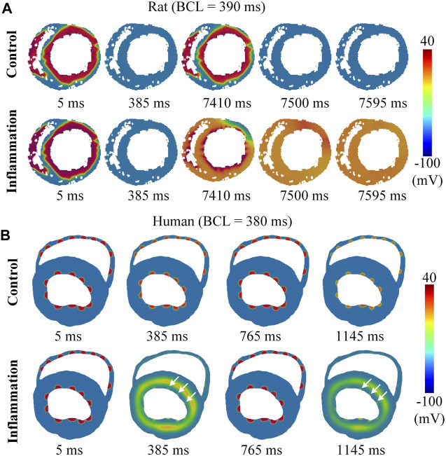
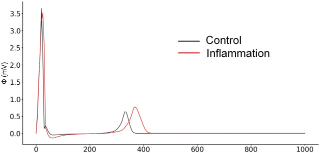
Cardiovascular diseases are the primary cause of death of humans, and among these, ventricular arrhythmias are the most common cause of death. There is plausible evidence implicating inflammation in the etiology of ventricular fibrillation (VF). In the case of systemic inflammation caused by an overactive immune response, the induced inflammatory cytokines directly affect the function of ion channels in cardiomyocytes, leading to a prolonged action potential duration (APD). However, the mechanistic links between inflammatory cytokine-induced molecular and cellular influences and inflammation-associated ventricular arrhythmias need to be elucidated. The present study aimed to determine the potential impact of systemic inflammation on ventricular electrophysiology by means of multiscale virtual heart models. The experimental data on the ionic current of three major cytokines [i.e., tumor necrosis factor-α (TNF-α), interleukin-1 (IL-1β), and interleukin-6 (IL-6)] were incorporated into the cell model, and the effects of each cytokine and their combined effect on the cell action potential (AP) were evaluated. Moreover, the integral effect of these cytokines on the conduction of excitation waves was also investigated in a tissue model. The simulation results suggested that inflammatory cytokines significantly prolonged APD, enhanced the transmural and regional repolarization heterogeneities that predispose to arrhythmias, and reduced the adaptability of ventricular tissue to fast heart rates. In addition, simulated pseudo-ECGs showed a prolonged QT interval-a manifestation consistent with clinical observations. In summary, the present study provides new insights into ventricular arrhythmias associated with inflammation.
Keywords: COVID-19; cardiac simulation; inflammation; rat ventricle; ventricular arrhythmia.
Copyright © 2022 Bi, Zhang, Jiang, Ma, Li, Lu, Yang and Wei.
Conflict of interest statement
The authors declare that the research was conducted in the absence of any commercial or financial relationships that could be construed as a potential conflict of interest.
Figures
Similar articles
-
Proarrhythmic effects of carbon monoxide in human ventricular tissue: insights from computational modeling.Comput Biol Med. 2022 Jan;140:105066. doi: 10.1016/j.compbiomed.2021.105066. Epub 2021 Nov 23. Comput Biol Med. 2022. PMID: 34839185
-
In silico investigation of pro-arrhythmic effects of azithromycin on the human ventricle.Biochem Biophys Rep. 2021 Jun 14;27:101043. doi: 10.1016/j.bbrep.2021.101043. eCollection 2021 Sep. Biochem Biophys Rep. 2021. PMID: 34179514 Free PMC article.
-
Effects of amiodarone on short QT syndrome variant 3 in human ventricles: a simulation study.Biomed Eng Online. 2017 Jun 7;16(1):69. doi: 10.1186/s12938-017-0369-0. Biomed Eng Online. 2017. PMID: 28592292 Free PMC article.
-
Antiviral and anti-inflammatory drugs to combat COVID-19: Effects on cardiac ion channels and risk of ventricular arrhythmias.Bioimpacts. 2022;12(1):9-20. doi: 10.34172/bi.2021.23630. Epub 2021 Dec 22. Bioimpacts. 2022. PMID: 35087712 Free PMC article. Review.
-
Review: The Prolonged QT Interval: Role of Pro-inflammatory Cytokines, Reactive Oxygen Species and the Ceramide and Sphingosine-1 Phosphate Pathways.In Vivo. 2015 Nov-Dec;29(6):619-36. In Vivo. 2015. PMID: 26546519 Review.
Cited by
-
In silico mechanisms of arsenic trioxide-induced cardiotoxicity.Front Physiol. 2022 Dec 15;13:1004605. doi: 10.3389/fphys.2022.1004605. eCollection 2022. Front Physiol. 2022. PMID: 36589437 Free PMC article.
-
What Do We Know So Far About Ventricular Arrhythmias and Sudden Cardiac Death Prediction in the Mitral Valve Prolapse Population? Could Biomarkers Help Us Predict Their Occurrence?Curr Cardiol Rep. 2024 May;26(5):245-268. doi: 10.1007/s11886-024-02030-9. Epub 2024 Mar 20. Curr Cardiol Rep. 2024. PMID: 38507154 Free PMC article. Review.
-
Computational analysis of long QT syndrome type 2 and the therapeutic effects of KCNQ1 antibodies.Digit Health. 2024 Oct 29;10:20552076241277032. doi: 10.1177/20552076241277032. eCollection 2024 Jan-Dec. Digit Health. 2024. PMID: 39484649 Free PMC article.
-
Stretch of the papillary insertion triggers reentrant arrhythmia: an in silico patient study.Front Physiol. 2024 Aug 19;15:1447938. doi: 10.3389/fphys.2024.1447938. eCollection 2024. Front Physiol. 2024. PMID: 39224207 Free PMC article.
-
COVID-19 and Cardiac Arrhythmias: Lesson Learned and Dilemmas.J Clin Med. 2024 Nov 29;13(23):7259. doi: 10.3390/jcm13237259. J Clin Med. 2024. PMID: 39685718 Free PMC article.
References
LinkOut - more resources
Full Text Sources