Assessment of Left Ventricular Systolic Function by Cardiovascular Magnetic Resonance Compressed Sensing Real-Time Cine Imaging Combined With Area-Length Method in Normal Sinus Rhythm and Atrial Fibrillation
- PMID: 35711346
- PMCID: PMC9197321
- DOI: 10.3389/fcvm.2022.896816
Assessment of Left Ventricular Systolic Function by Cardiovascular Magnetic Resonance Compressed Sensing Real-Time Cine Imaging Combined With Area-Length Method in Normal Sinus Rhythm and Atrial Fibrillation
Abstract
Background: The most-commonly used multi-slice Simpson's method employed with routine two-dimensional segmented cine images makes it difficult to evaluate left ventricular (LV) volume and function due to endocardial border blurring and beat-to-beat variation during atrial fibrillation (AF) status.
Objectives: To assess the feasibility of compressed sensing real-time (CSRT) cine imaging combined with an area-length method for quantification of LV systolic function in normal sinus rhythm (NSR) and AF.
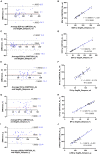
Methods: The CSRT cine sequence and routine segmented balanced Steady-State-Free-Precession cine sequence were performed in 71 patients with NSR (n = 36) or AF (n = 35). Image quality and edge sharpness for both sequences were assessed. The LV functional measurements in patients with NSR included end-diastolic volume (EDV), end-systolic volume (ESV), stroke volume (SV), ejection fraction (EF), cardiac output (CO), cardiac index (CI), and LV mass (LVM); all were assessed using segmented cine with Simpson's rule in short axis (SegSA_Simpson, as a reference standard) and area-length (AL) method in the two chamber (Seg2CH_AL) or four chamber (Seg4CH_AL) and CSRT cine with AL method in the two chamber (CSRT2CH_AL) or four chamber (CSRT4CH_AL). Finally, the mean, maximum, and minimum values of each LV functional parameter [EDV/ESV/SV/EF/CO/CI/LVM/heart rate (HR)] from 4~5 consecutive heartbeats were measured using CSRT2CH_AL in patients with AF.
Results: In patients with NSR, measurements of EDV (p > 0.05), ESV (p > 0.05), SV (p > 0.05), EF (p > 0.05), and LVM (p > 0.05) assessed with CSRT2CH_AL did not differ significantly from those obtained with SegSA_Simpson. In patients with AF, CSRT image quality score (p < 0.001) and edge sharpness (p < 0.001) both were significantly higher than those obtained from segmented cine. The CSRT2CH_AL provided significantly different results among mean, maximum, and minimum values of each LV parameter from 4~5 consecutive heartbeats (all p < 0.001) with strong inter- and intra-observer agreement in AF.
Conclusions: The CSRT cine sequence combined with two chamber area-length analysis accurately assessed LV systolic function in NSR. This approach is expected to permit the assessment of multiple parameters in consecutive heartbeats with good inter- and intra-observer reproducibility for beat-to-beat analysis of LV function in AF.
Keywords: area-length; atrial fibrillation; cardiovascular magnetic resonance; compressed sensing real-time; normal sinus rhythm.
Copyright © 2022 Yin, Cui, An, Zhao, Yang, Li, Yang, Wang, Dong, Yu, He, Chen, Lu and Zhao.
Conflict of interest statement
JA was employed by Siemens Shenzhen Magnetic Resonance Ltd. The remaining authors declare that the research was conducted in the absence of any commercial or financial relationships that could be construed as a potential conflict of interest.
Figures


Similar articles
-
Feasibility, accuracy, and reproducibility of real-time full-volume 3D transthoracic echocardiography to measure LV volumes and systolic function: a fully automated endocardial contouring algorithm in sinus rhythm and atrial fibrillation.JACC Cardiovasc Imaging. 2012 Mar;5(3):239-51. doi: 10.1016/j.jcmg.2011.12.012. JACC Cardiovasc Imaging. 2012. PMID: 22421168
-
[Feasibility of Single-Breath-Hold Compressed Sensing Real-Time Cine Imaging for Assessment of Ventricular Function and Left Ventricular Strain in Cardiac Magnetic Resonance].Sichuan Da Xue Xue Bao Yi Xue Ban. 2022 May;53(3):497-503. doi: 10.12182/20220560506. Sichuan Da Xue Xue Bao Yi Xue Ban. 2022. PMID: 35642161 Free PMC article. Chinese.
-
Compressed sensing real-time cine cardiovascular magnetic resonance: accurate assessment of left ventricular function in a single-breath-hold.J Cardiovasc Magn Reson. 2016 Aug 24;18(1):50. doi: 10.1186/s12968-016-0271-0. J Cardiovasc Magn Reson. 2016. PMID: 27553656 Free PMC article.
-
Cardiac cine with compressed sensing real-time imaging and retrospective motion correction for free-breathing assessment of left ventricular function and strain in clinical practice.Quant Imaging Med Surg. 2023 Apr 1;13(4):2262-2277. doi: 10.21037/qims-22-596. Epub 2023 Mar 1. Quant Imaging Med Surg. 2023. PMID: 37064398 Free PMC article.
-
Imaging of arrhythmia: Real-time cardiac magnetic resonance imaging in atrial fibrillation.Eur J Radiol Open. 2022 Mar 2;9:100404. doi: 10.1016/j.ejro.2022.100404. eCollection 2022. Eur J Radiol Open. 2022. PMID: 35265735 Free PMC article.
Cited by
-
Case Report: Hemodynamic consequences of severe supraventricular arrhythmia assessed by real-time magnetic resonance imaging in combination with electrocardiography.Front Cardiovasc Med. 2025 Jun 23;12:1596440. doi: 10.3389/fcvm.2025.1596440. eCollection 2025. Front Cardiovasc Med. 2025. PMID: 40625394 Free PMC article.
-
Comparing Strain Assessment in Compressed Sensing and Conventional Cine MRI.J Imaging Inform Med. 2024 Aug;37(4):1933-1943. doi: 10.1007/s10278-024-01040-x. Epub 2024 Feb 22. J Imaging Inform Med. 2024. PMID: 38388867 Free PMC article.
-
ECG-free cine MRI with data-driven clustering of cardiac motion for quantification of ventricular function.NMR Biomed. 2024 Apr;37(4):e5091. doi: 10.1002/nbm.5091. Epub 2024 Jan 9. NMR Biomed. 2024. PMID: 38196195 Free PMC article.
-
Dynamic Regularized Adaptive Cluster Optimization (DRACO) for Quantitative Cardiac Cine MRI in Complex Arrhythmias.J Magn Reson Imaging. 2025 Jan;61(1):248-262. doi: 10.1002/jmri.29425. Epub 2024 May 6. J Magn Reson Imaging. 2025. PMID: 38708951
References
-
- Grothues F, Smith GC, Moon JC, Bellenger NG, Collins P, Klein HU, et al. . Comparison of interstudy reproducibility of cardiovascular magnetic resonance with two-dimensional echocardiography in normal subjects and in patients with heart failure or left ventricular hypertrophy. Am J Cardiol. (2002) 90:29–34. 10.1016/S0002-9149(02)02381-0 - DOI - PubMed
-
- Thiele H, Paetsch I, Schnackenburg B, Bornstedt A, Grebe O, Wellnhofer E, et al. . Improved accuracy of quantitative assessment of left ventricular volume and ejection fraction by geometric models with steady-state free precession. J Cardiovasc Magn Reson. (2002) 4:327–39. 10.1081/JCMR-120013298 - DOI - PubMed
-
- Hazirolan T, Taşbaş B, Dagoglu MG, Canyigit M, Abali G, Aytemir K, et al. . Comparison of short and long axis methods in cardiac MR imaging and echocardiography for left ventricular function. Diagn Interv Radiol (Ankara, Turkey). (2007) 13:33–8. - PubMed
LinkOut - more resources
Full Text Sources

