Review
. 2022 May 30:9:869731.
doi: 10.3389/fsurg.2022.869731. eCollection 2022.

The Preperitoneal Space in Hernia Repair

Affiliations
Review

The Preperitoneal Space in Hernia Repair

A Lorenz et al. Front Surg. .

Abstract

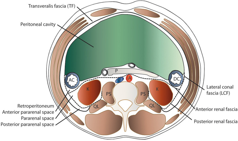
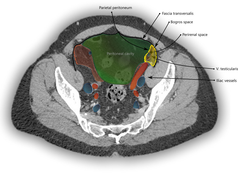
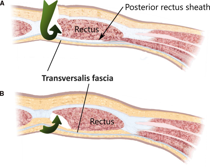
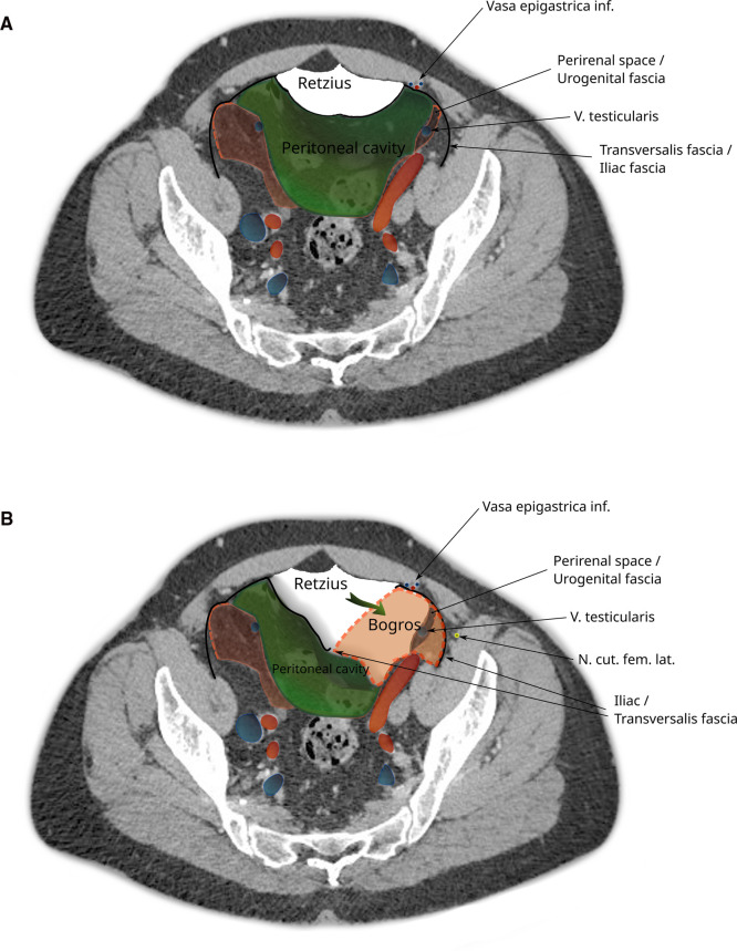
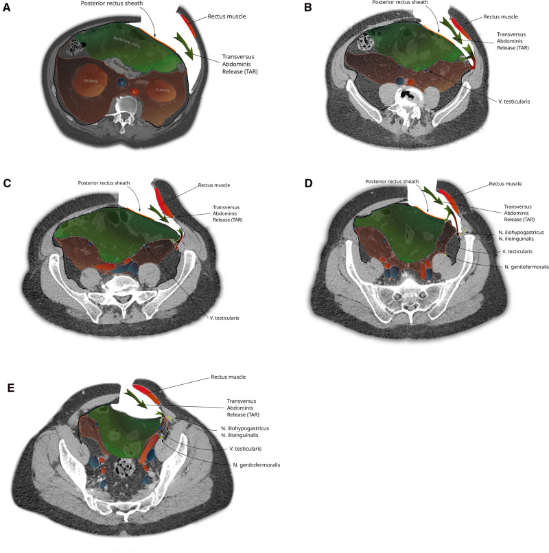
The preperitoneal spaces relevant for incisional hernia repair and minimally invasive groin hernia repair are described in terms of surgical anatomy. Emphasis is put on the transversalis fascia and the urogenital fascia and its extensions, the vesicoumbilical fascia, and the spermatic sheath of Stoppa procedure. Steps in hernia surgery where these structures are relevant are reviewed.

Keywords: abdominal wall repair; anatomy; groin hernia; retroperitoneum; transversalis fascia.


Conflict of interest statement

The authors declare that the research was conducted in the absence of any commercial or financial relationships that could be construed as a potential conflict of interest.

Figures

Figure 1
Figure 1
Schematic transverse section of the abdomen with emphasis on the extraperitoneal spaces. The abdominal structures are separated from the musculoskeletal structures by the transversalis fascia (TF). The peritoneal cavity is delimited by the parietal peritoneum, which is the anterior border of the anterior pararenal space (APS) that contains the ascending (AC) and descending (DC) colon, pancreas (P), and duodenum (D). Posteriorly lies the perirenal space (PRS) with the kidney (K) and medially the aorta (A) and vena cava (V), delimited by the anterior renal fascia (anterior RF or Gerota’s fascia) and the posterior renal fascia (posterior RF or Zuckerkandl’s) and laterally the lateroconal fascia (LCF). Dorsally, the quadratus lumborum (QL) and psoas major (PS) muscles are found within the spine.
Figure 2
Figure 2
Computed tomography imaging with a reconstruction of the transverse and sagittal planes with contrast enhancement (A) and emphasizing the perirenal fat pad and its limits. (B) Same images with the posterior renal fascia highlighted in blue, the anterior renal fascia in red, and TF in black.
Figure 3
Figure 3
CT imaging with a transverse section through the pelvis at the apex of the urinary bladder. The space of Bogros is marked in yellow and limited by the TF (black) laterally and the parietal peritoneum (green) medially. It contains the extension of the perirenal space (red) into the pelvis.
Figure 4
Figure 4
The surgical access (green arrows) of a total extraperitonal hernioplasty through the layers of the ventral abdominal wall are depicted (A) cranial and (B) caudal of the arcuate area. The transversalis fascia passes the posterior rectus sheath dorsally and continues beyond the arcuate area and separates the musculoaponeurotic structures from the visceral peritoneum.
Figure 5
Figure 5
CT image of the pelvis showing dissection during a total extraperitoneal hernioplasty (TEP). (A) The peritoneal sac is displaced and the space of Retzius dissected. (B) The space of Bogros is opened by cleaving the transversalis fascia. Note that the space of Bogros contains the spermatic vessels and tissues from the perirenal space enveloped by the urogenital fascia that extends from the abdomen into the pelvis.
Figure 6
Figure 6
The steps of a transversus abdominis release-maneuver (TAR): (A) Lateral incision of the posterior rectus sheath and dorsolateral dissection along the transverse abdominis muscle (green arrow). Caudal extension of the dissection follows the urogenital fascia into the pelvis (B-E) reaching the space of Bogros in the superficial parietal layer that is so carefully avoided during a TEP procedure (red part of bifurcated arrow). A switch of dissection into the deep visceral layer, analogue to the dissection in TEP is necessary if a prosthetic mesh should also cover a groin hernia (green part of bifurcated arrow).
Figure 7
Figure 7
CT imaging with coronal (A) and sagittal (B) sections through the abdomen. The pararenal fat extends into the pelvis and in this patient ultimately into the left inguinal canal like an inverted cone. An attachment of the posterior renal fascia to the transversalis fascia at the ililac crest can be seen in the sagittal section (*) and (C) transverse section (arrows).

References

    1. Cooper AP. The anatomy and surgical treatment of inguinal and congential hernia. London, United Kingdom (1804).
    1. Cruveilhier J. The anatomy of the human body. New York: Harper & Brothers; (1844). 305 p.
    1. Mackay JY. The relations of the aponeurosis of the transversalis and internal oblique muscles to the deep epigastric artery and to the inguinal canal. In: Cleland J, Mackay JY, Young RB, editors. Memoirs and memoranda in anatomy, Vol. I. London: Williams and Norgate; (1889). p. 143–5.
    1. McVay C, Anson BJ. Composition of the rectus sheath. Anat Rec. (1940) 77:213–25. 10.1002/ar.1090770210 - DOI
    1. Lytle WJ. The internal inguinal ring. Br J Surg. (1945) 32:441–6. 10.1002/bjs.18003212802 - DOI

LinkOut - more resources