. 2022 May 30:13:884210.
doi: 10.3389/fgene.2022.884210. eCollection 2022.

The Pathogenic R3052W BRCA2 Variant Disrupts Homology-Directed Repair by Failing to Localize to the Nucleus

Affiliations

The Pathogenic R3052W BRCA2 Variant Disrupts Homology-Directed Repair by Failing to Localize to the Nucleus

Judit Jimenez-Sainz et al. Front Genet. .

Abstract

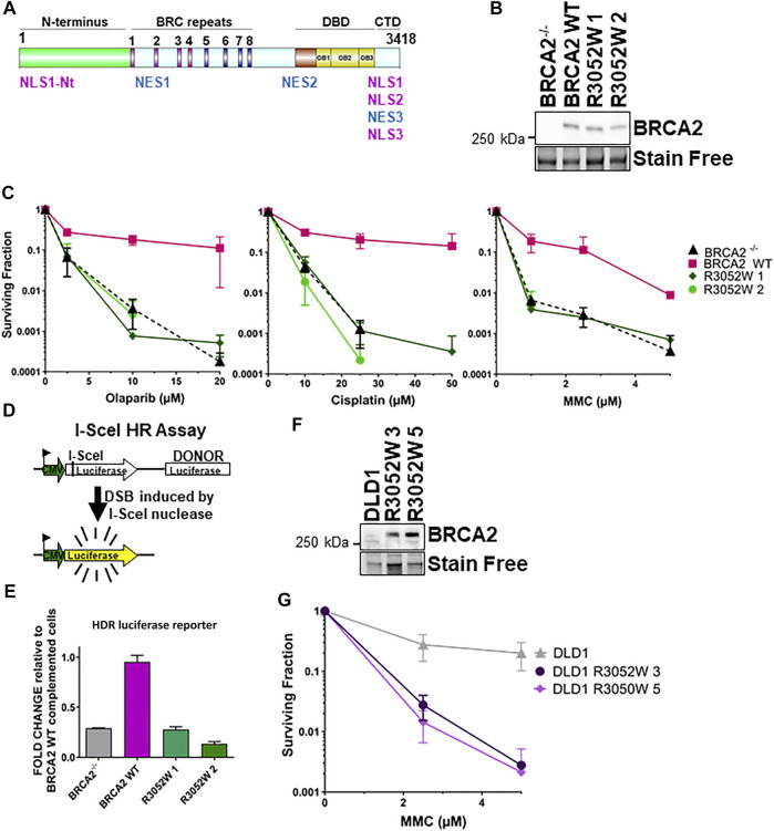
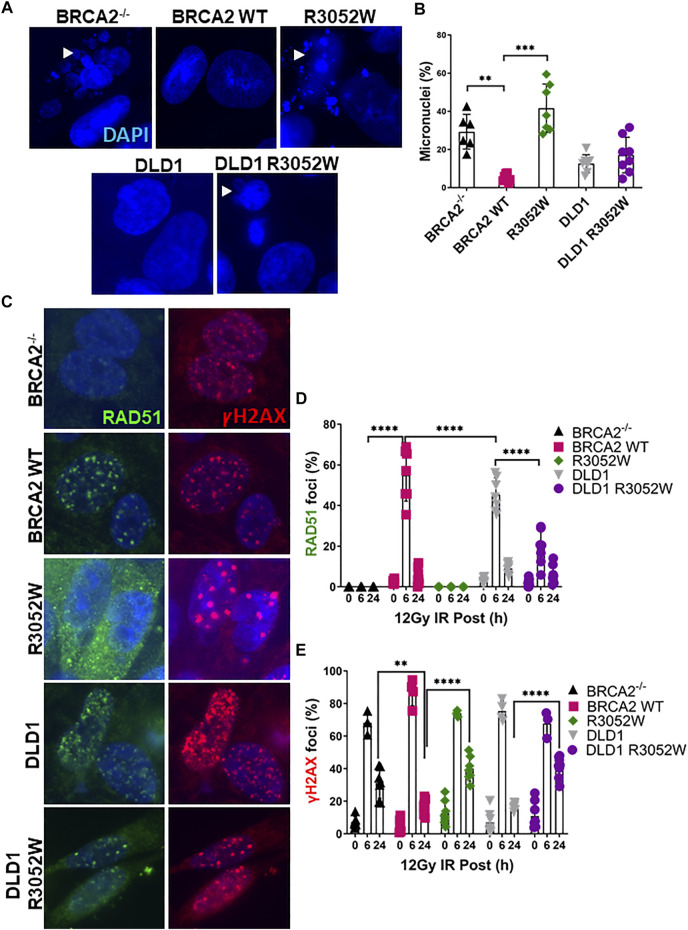
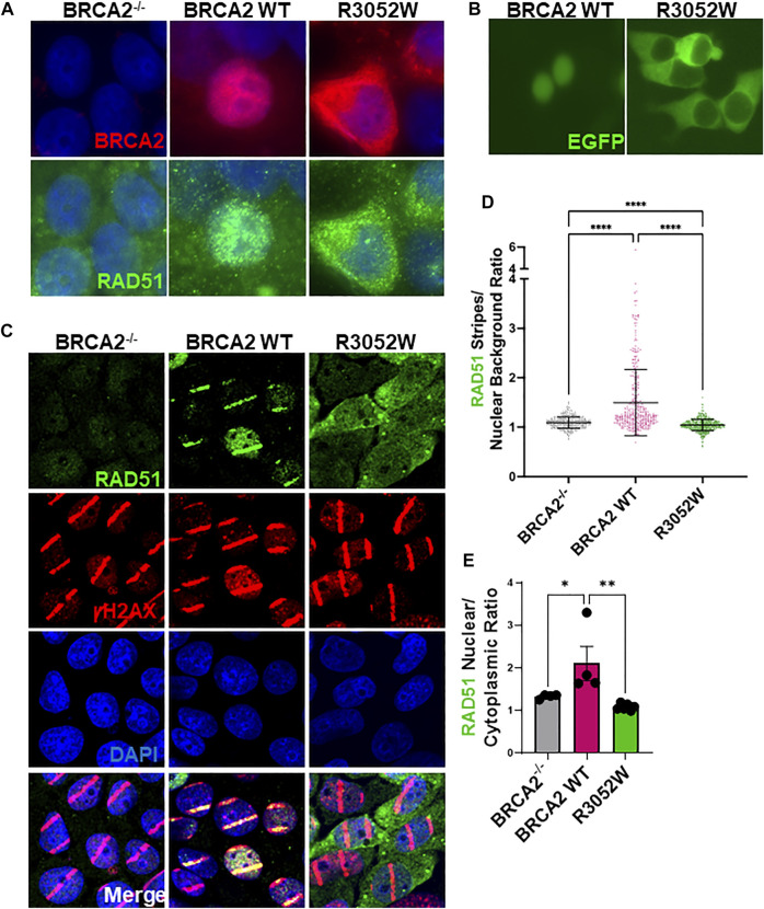
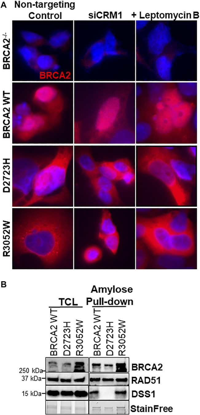
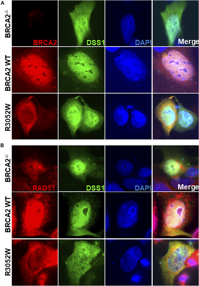
The BRCA2 germline missense variant, R3052W, resides in the DNA binding domain and has been previously classified as a pathogenic allele. In this study, we sought to determine how R3052W alters the cellular functions of BRCA2 in the DNA damage response. The BRCA2 R3052W mutated protein exacerbates genome instability, is unable to rescue homology-directed repair, and fails to complement cell survival following exposure to PARP inhibitors and crosslinking drugs. Surprisingly, despite anticipated defects in DNA binding or RAD51-mediated DNA strand exchange, the BRCA2 R3052W protein mislocalizes to the cytoplasm precluding its ability to perform any DNA repair functions. Rather than acting as a simple loss-of-function mutation, R3052W behaves as a dominant negative allele, likely by sequestering RAD51 in the cytoplasm.

Keywords: BRCA2; DNA repair; DSS1; R3052W; RAD51; homology-directed repair; nuclear localization.


Conflict of interest statement

The authors declare that the research was conducted in the absence of any commercial or financial relationships that could be construed as a potential conflict of interest.

Figures

FIGURE 1
FIGURE 1
The BRCA2 R3052W mutation fails to complement chemotherapeutic sensitivity and homology-directed repair functions in BRCA2 knockout cells. Overexpression of R3052W in DLD1 parental BRCA2 wild-type cells confers sensitivity to MMC DNA damage. (A) BRCA2 protein schematic depicting domain organization: N-terminus, BRC repeats DNA binding domain (DBD), and C-terminal domain (CTD). BRCA2 nuclear localization and export sequences are listed. NLS1-Nt (433–436 aa), NLS1 (3,265–3,270 aa) NLS2 (3,311–3,317 aa), NLS3 (3,381–3,385 aa), NES1 (1,383–1,392 aa), NES2 (2,682–2,698 aa) and NES3 (3,270–3,280 aa). (B) Western blot of total cellular lysates from DLD-1 BRCA2−/− cells stably transfected with full-length BRCA2 cDNA constructs: BRCA2 Wild Type (WT) and BRCA2 R3052W (1 and 2 correspond to two independent clones). BRCA2 cDNAs contain a 2XMBP tag on the N-terminus and were detected with an MBP antibody. 2XMBP-BRCA2 (470 kDa). Stain Free is a protein-loading control. (C) Clonogenic survival analyses of BRCA2 WT versus the two independent R3052W clones after treatment with Olaparib, cisplatin, and mitomycin C. (D) Schematic of I-SceI nuclease-induced DSB HDR luciferase assay. (E) Quantification of luciferase activity (normalized to BRCA2 WT as 1). Error bars are SD (n = 5). (F) Western blot of total cellular lysates from DLD-1 parental cells (these cells express a wild-type allele of BRCA2) stably transfected with R3052W (3 and 5 correspond to two independent clones) full-length 2XMBP-BRCA2 cDNA constructs. BRCA2 was detected with an MBP antibody. (G) Clonogenic survival analyses of DLD parental clones upon treatment with mitomycin C.
FIGURE 2
FIGURE 2
The BRCA2 R3052W mutation exacerbates micronuclei formation and interferes with RAD51 foci formation in DLD1 parental and BRCA2 wild-type cells. (A) Representative immunofluorescent images of micronuclei (blue) in a DLD1 BRCA2 knockout cell line (top) expressing WT or the R3052W mutant and parental DLD1 cells (bottom) compared to a stable cell line expressing the R3052W protein. (B) Quantification of the percentage of cells with micronuclei. (C) Representative immunofluorescent images of RAD51 (green) and gammaH2AX (red) foci, and DAPI staining to visualize nuclei (blue). Cells were fixed and imaged 6 h post-IR (12 Gy). (D) Quantification of the percentage of cells with RAD51 foci. (E) Quantification of the percentage of cells with gammaH2AX. (Quantification represents 3 independent experiments and statistical analysis t-test and one-way ANOVA. **p-value < 0.01, ***p value < 0.001, ****p value < 0.0001).
FIGURE 3
FIGURE 3
The R3052W protein localizes to the cytosol. (A) Immunofluorescent localization of BRCA2 in untreated BRCA2 knockout cells (BRCA2−/−), and stable cell lines expressing either BRCA2 WT or R3052W. Representative images of 2XMBP-BRCA2 (red, anti-MBP), RAD51 (green), and nuclei (blue). (B) Live images of BRCA2 knockout cells expressing either BRCA2 WT or R3052W fused to GFP at the N-terminus. (C) Representative immunofluorescence images of laser micro-irradiation experiments in BRCA2 knockout cells stably expressing BRCA2 WT or R3052W. DNA damage (stripes) are depicted in red (gammaH2AX), green (RAD51), and nuclei in blue (DAPI). (D) Quantification of RAD51 fluorescence intensity in damage areas (stripes) over the background in non-irradiated areas of respective laser micro-irradiated nuclei. Each data point represents a single analyzed nucleus, while the solid line is a mean value ± SD (Kruskal-Wallis test with Dunn’s multiple comparison post hoc test; **** p value < 0.0001). (E) Quantification of RAD51 intensity in the nuclear and cytoplasmic compartments. Each data point represents a single analyzed area (220.16 microns × 220.16 microns), while bars represent mean ± SD (one-way ANOVA with Holm-Šídák’s multiple comparisons post hoc test, * p-value<0.05; ** p-value<0.01).
FIGURE 4
FIGURE 4
R3052W cytoplasmic localization is not altered by CRM1 depletion or leptomycin treatment and retains binding to DSS1. (A) Immunofluorescent localization of BRCA2 in untreated stable cell lines expressing WT, D2723H, or R3052W BRCA2 proteins upon RNA interference-mediated depletion of CRM1 or treatment with the nuclear export inhibitor leptomycin B. Representative images of BRCA2 (red, MBP antibody) and DAPI staining to visualize nuclei (blue). (B) Western blots of total cellular lysates (TCL) and amylose pulldowns from HEK 293T cells transiently transfected with 2XMBP-BRCA2 WT, D2723H, or R3052W co-transfected with HA-DSS1. Anti-MBP antibody was used for BRCA2 detection, Anti-RAD51 antibody was used for endogenous RAD51 detection and Anti-HA antibody was used for DSS1 detection.
FIGURE 5
FIGURE 5
Ectopic expression of DSS1 does not alter BRCA2 WT, R3052W or RAD51 cellular localization. (A) Immunofluorescent localization of BRCA2 and DSS1 in untreated BRCA2 knockout cells stably expressing WT BRCA2 or the R3052W mutant concurrent with transient expression of HA-DSS1. Representative images of BRCA2 (red, MBP antibody), DSS1 (green, HA antibody) and DAPI staining to visualize nuclei (blue). (B) Immunofluorescent localization of RAD51 and DSS1 in untreated BRCA2 knockout cells stably expressing WT BRCA2 or the R3052W mutant concurrent with transient expression of HA-DSS1. Representative images of RAD51 (red, RAD51 antibody), DSS1 (green, HA antibody), and DAPI staining to visualize nuclei (blue).

Similar articles

Cited by

References

    1. Ban S., Shinohara T., Hirai Y., Moritaku Y., Cologne J. B., Macphee D. G. (2001). Chromosomal Instability in BRCA1- or BRCA2-Defective Human Cancer Cells Detected by Spontaneous Micronucleus Assay. Mutat Res. 474, 15–23. - PubMed
    1. Bryant H. E., Schultz N., Thomas H. D., Parker K. M., Flower D., Lopez E., et al. (2005). Specific Killing of BRCA2-Deficient Tumours with Inhibitors of poly(ADP-Ribose) Polymerase. Nature 434, 913–917. 10.1038/nature03443 - DOI - PubMed
    1. Capanu M., Concannon P., Haile R. W., Bernstein L., Malone K. E., Lynch C. F., et al. (2011). Assessment of Rare BRCA1 and BRCA2 Variants of Unknown Significance Using Hierarchical Modeling. Genet. Epidemiol. 35, 389–397. 10.1002/gepi.20587 - DOI - PMC - PubMed
    1. Chatterjee G., Jimenez-Sainz J., Presti T., Nguyen T., Jensen R. B. (2016). Distinct Binding of BRCA2 BRC Repeats to RAD51 Generates Differential DNA Damage Sensitivity. Nucleic Acids Res. 44, 5256–5270. 10.1093/nar/gkw242 - DOI - PMC - PubMed
    1. Cunningham J. M., Cicek M. S., Larson N. B., Davila J., Wang C., Larson M. C., et al. (2014). Clinical Characteristics of Ovarian Cancer Classified by BRCA1, BRCA2 and RAD51C Status. Sci. Rep. 4, 4026. 10.1038/srep04026 - DOI - PMC - PubMed