[Effect of M2-like macrophage/microglia-derived mitochondria transplantation in treatment of mouse spinal cord injury]
- PMID: 35712934
- PMCID: PMC9240838
- DOI: 10.7507/1002-1892.202201040
[Effect of M2-like macrophage/microglia-derived mitochondria transplantation in treatment of mouse spinal cord injury]
Abstract
Objective: To investigate the effect of M2-like macrophage/microglia-derived mitochondria transplantation in treatment of mouse spinal cord injury (SCI).
Methods: BV2 cells were classified into M1 (LPS treatment), M2 (IL-4 treatment), and M0 (no treatment) groups. After receiving M1 and M2 polarization, BV2 cells received microscopic observation, immunofluorescence staining [Arginase-1 (Arg-1)] and flow cytometry [inducible nitric oxide synthase (iNOS), Arg-1] to determine the result of polarization. MitoSox Red and 2, 7-dichlorodi-hydrofluorescein diacetate (DCFH-DA) stainings were used to evaluate mitochondrial function difference. Mitochondria was isolated from M2-like BV2 cells through differential velocity centrifugation for following transplantation. Then Western blot was used to measure the expression levels of the relevant complexes (complexes Ⅱ, Ⅲ, Ⅳ, and Ⅴ) in the oxidative phosphorylation (OXPHOS), and compared with M2-like BV2 cells to evaluate whether the mitochondria were obtained. Thirty-six female C57BL/6 mice were randomly divided into 3 groups ( n=12). Mice from sham group were only received the T 10 laminectomy. After the T 10 spinal cord injury (SCI) model was prepared in the SCI group and mitochondria transplantation (MT) group, mitochondrial storage solution and mitochondria (100 μg) derived from M2-like BV2 cells were injected into the injured segment, respectively. After operation, the Basso Mouse Scale (BMS) score was performed to evaluate the motor function recovery. And immunofluorescence staining, lycopersicon esculentum agglutinin (LEA)-FITC staining, and ELISA [vascular endothelial growth factor A (VEGFA)] were also performed.
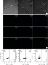
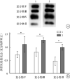
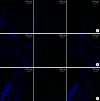
Results: After polarization induction, BV2 cells in M1 and M2 groups showed specific morphological changes of M1-like and M2-like macrophages, respectively. Immunofluorescence staining showed that the positive expression of M2-like macrophages marker (Arg-1) was significantly higher in M2 group than in M0 group and M1 group ( P<0.05). Flow cytometry showed that the expression of M1-like macrophage marker (iNOS) was significantly higher in M1 group than in M0 group and M2 group ( P<0.05), and the expression of Arg-1 was significantly higher in M2 group than in M0 group and M1 group ( P<0.05). MitoSox Red and DCFH-DA stainings showed that the fluorescence intensity of the M2 group was significantly lower than that of the M1 group ( P<0.05), and there was no significant difference with the M0 group ( P>0.05). The M2-like BV2 cells-derived mitochondria was identified through Western blot assay. Animal experiments showed that the BMS scores of MT group at 21 and 28 days after operation were significantly higher than those of SCI group ( P<0.05). At 14 days after operation, the number of iNOS-positive cells in MT group was significantly lower than that in SCI group ( P<0.05), but still higher than that in sham group ( P<0.05); the number of LEA-positive cells and the expression of VEGFA in MT group were significantly more than those in the other two groups ( P<0.05).
Conclusion: M2-like macrophage/microglia-derived mitochondria transplantation can promote angiogenesis and inhibit inflammatory M1-like macrophage/microglia polarization after mouse SCI to improve function recovery.
目的: 探讨M2型巨噬细胞/小胶质细胞来源线粒体移植治疗小鼠脊髓损伤的效果。.
方法: 取小鼠小胶质细胞(BV2细胞)分别采用脂多糖(lipopolysaccharide,LPS)、IL-4诱导其向M1型(M1组)及M2型(M2组)极化,以未作任何干预的细胞作为对照(M0组);光镜下观察细胞形态,免疫荧光染色 [精氨酸酶 1(Arginase 1,Arg-1)] 及流式细胞术 [诱导型一氧化氮合酶(inducible nitric oxide synthase,iNOS)、Arg-1] 鉴定极化后细胞类型,并通过MitoSox Red与DCFH-DA染色评估细胞线粒体功能。使用差速离心法提取M2组BV2细胞来源线粒体,Western blot法测量氧化磷酸酶链(oxidative phosphorylation,OXPHOS)内各相关复合物(复合物Ⅱ、Ⅲ、Ⅳ与Ⅴ)的表达水平,并与M2型BV2细胞比较,以评估是否获得所需线粒体。取36只雌性C57BL/6小鼠随机分为3组( n=12),其中假手术组仅切除T 10椎板,脊髓损伤组及线粒体移植组制备T 10脊髓损伤模型后,于损伤节段分别注射线粒体储存液及M2型BV2细胞来源线粒体(100 μg)。术后采用BMS(Basso Mouse Scale)评分评价小鼠后肢运动功能,取材行免疫荧光染色(iNOS)、LEA-FITC染色及ELISA检测(VEGFA)。.
结果: 极化诱导后,M1组及M2组BV2细胞分别呈现M1型及M2型巨噬细胞特异性形态改变;免疫荧光染色示M2组的M2型巨噬细胞标志物Arg-1染色阳性表达高于M0组及M1组( P<0.05);流式细胞术检测示M1组的M1型巨噬细胞标志物iNOS表达高于M0组及M2组( P<0.05),M2组Arg-1表达高于M0及M1组( P<0.05)。MitoSox Red及DCFH-DA染色示M2组荧光强度均低于M1组( P<0.05),与M0组差异无统计学意义( P>0.05)。Western blot法鉴定成功提取M2型BV2细胞来源线粒体。动物实验示,线粒体移植组术后21、28 d BMS评分高于脊髓损伤组( P<0.05);术后14 d损伤局部iNOS阳性细胞数低于脊髓损伤组( P<0.05),但仍高于假手术组( P<0.05); LEA阳性细胞较其余两组增多;VEGFA表达亦高于其余两组( P<0.05)。.
结论: M2型巨噬细胞/小胶质细胞来源线粒体可通过促进损伤局部血管新生并抑制炎症性M1型巨噬细胞极化,促进小鼠脊髓损伤修复。.
Keywords: Spinal cord injury; angiogenesis; macrophage; mice; microglia; mitochondria transplantation.
Conflict of interest statement
利益冲突 在课题研究和文章撰写过程中不存在利益冲突;经费支持没有影响文章观点和对研究数据客观结果的统计分析及其报道
Figures


Similar articles
-
Mitochondrial-targeting antioxidant MitoQ modulates angiogenesis and promotes functional recovery after spinal cord injury.Brain Res. 2022 Jul 1;1786:147902. doi: 10.1016/j.brainres.2022.147902. Epub 2022 Apr 4. Brain Res. 2022. PMID: 35381215
-
[Experimental study of M2 microglia transplantation promoting spinal cord injury repair in mice].Zhongguo Xiu Fu Chong Jian Wai Ke Za Zhi. 2024 Feb 15;38(2):198-205. doi: 10.7507/1002-1892.202311093. Zhongguo Xiu Fu Chong Jian Wai Ke Za Zhi. 2024. PMID: 38385233 Free PMC article. Chinese.
-
[Necrostatin-1 promotes locomotor recovery after spinal cord injury through inhibiting apoptosis and M1 polarization of microglia/macrophage in mice].Xi Bao Yu Fen Zi Mian Yi Xue Za Zhi. 2021 Sep;37(9):775-780. Xi Bao Yu Fen Zi Mian Yi Xue Za Zhi. 2021. PMID: 34533123 Chinese.
-
Advances in the research of the role of macrophage/microglia polarization-mediated inflammatory response in spinal cord injury.Front Immunol. 2022 Dec 1;13:1014013. doi: 10.3389/fimmu.2022.1014013. eCollection 2022. Front Immunol. 2022. PMID: 36532022 Free PMC article.
-
The role and mechanisms of mesenchymal stem cells regulating macrophage plasticity in spinal cord injury.Biomed Pharmacother. 2023 Dec;168:115632. doi: 10.1016/j.biopha.2023.115632. Epub 2023 Oct 6. Biomed Pharmacother. 2023. PMID: 37806094 Review.
Cited by
-
Regulation of dynamic spatiotemporal inflammation by nanomaterials in spinal cord injury.J Nanobiotechnology. 2024 Dec 19;22(1):767. doi: 10.1186/s12951-024-03037-8. J Nanobiotechnology. 2024. PMID: 39696584 Free PMC article. Review.
-
Recent advances in mitochondrial transplantation to treat disease.Biomater Transl. 2025 Mar 25;6(1):4-23. doi: 10.12336/biomatertransl.2025.01.002. eCollection 2025. Biomater Transl. 2025. PMID: 40313574 Free PMC article. Review.
References
-
-
Rabchevsky AG, Michael FM, Patel SP. Mitochondria focused neurotherapeutics for spinal cord injury. Exp Neurol, 2020, 330: 113332. doi: 10.1016/j.expneurol.2020.113332.
-
-
-
Abbaszadeh F, Fakhri S, Khan H. Targeting apoptosis and autophagy following spinal cord injury: Therapeutic approaches to polyphenols and candidate phytochemicals. Pharmacol Res, 2020, 160: 105069. doi: 10.1016/j.phrs.2020.105069.
-
-
-
Simmons EC, Scholpa NE, Schnellmann RG. FDA-approved 5-HT 1F receptor agonist lasmiditan induces mitochondrial biogenesis and enhances locomotor and blood-spinal cord barrier recovery after spinal cord injury. Exp Neurol, 2021, 341: 113720. doi: 10.1016/j.expneurol.2021.113720.
-
MeSH terms
Substances
LinkOut - more resources
Full Text Sources
Medical
Research Materials
Miscellaneous
