. 2022 Jun 17;79(7):366.
doi: 10.1007/s00018-022-04387-y.

Mechanisms of chronic alcohol exposure-induced aggressiveness in cellular model of HCC and recovery after alcohol withdrawal

Affiliations

Mechanisms of chronic alcohol exposure-induced aggressiveness in cellular model of HCC and recovery after alcohol withdrawal

Constance Marié et al. Cell Mol Life Sci. .

Abstract

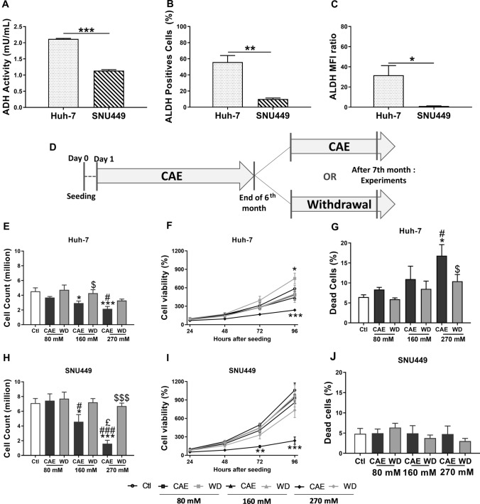
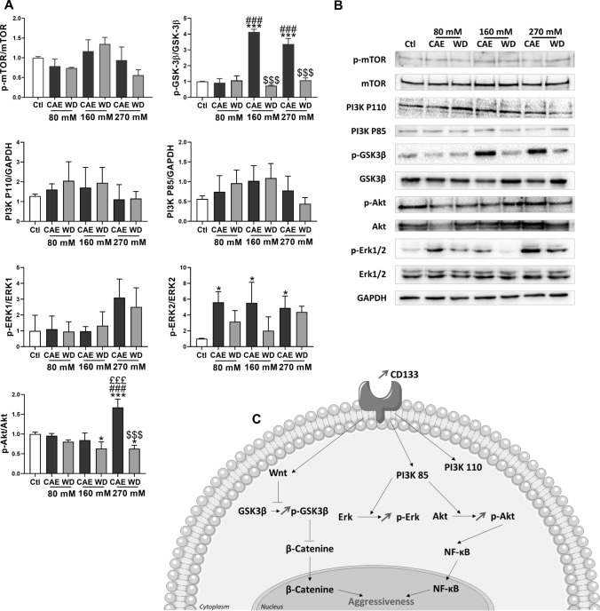
Alcohol-related liver disease is the most prevalent chronic liver disease worldwide, accounting for 30% of hepatocellular carcinoma (HCC) cases and HCC-specific deaths. However, the knowledge on mechanisms by which alcohol consumption leads to cancer progression and its aggressiveness is limited. Better understanding of the clinical features and the mechanisms of alcohol-induced HCC are of critical importance for prevention and the development of novel treatments. Early stage Huh-7 and advanced SNU449 liver cancer cell lines were subjected to chronic alcohol exposure (CAE), at different doses for 6 months followed by 1-month alcohol withdrawal period. ADH activity and ALDH expression were much lower in SNU449 compared with Huh-7 cells and at the 270 mM dose, CAE decreased cell viability by about 50% and 80%, respectively, in Huh-7 and SNU449 cells but induced mortality only in Huh-7 cells. Thus, Huh-7 may be more vulnerable to ethanol toxicity because of the higher levels of acetaldehyde. CAE induced a dose-dependent increase in cell migration and invasion and also in the expression of cancer stem cells markers (CD133, CD44, CD90). CAE in Huh-7 cells selectively activated ERK1/2 and inhibited GSK3β signaling pathways. Most of the changes induced by CAE were reversed after alcohol withdrawal. Interestingly, we confirmed the increase in CD133 mRNA levels in the tumoral tissue of patients with ethanol-related HCC compared to other HCC etiologies. Our results may explain the benefits observed in epidemiological studies showing a significant increase of overall survival in abstinent compared with non-abstinent patients.

Keywords: Alcohol; Cancerous stem cell; Liver cancer; Withdrawal.


Conflict of interest statement

No conflict of interest.

Figures

Fig. 1
Fig. 1
CAE protocol on HCC cell lines, alcohol metabolism, cell viability, and benefits of withdrawal. Cells were cultured in the presence of several doses of alcohol (80 mM, 160 mM or 270 mM) during 6 months. At the end of the 6th month, cells were splitted in two parts: one which continued its alcohol exposition, and the second was subject to a withdrawal during 1 month (A). Alcohol DeHydrogenase (ADH) activity was studied in Huh-7 and SNU449 cells. Results represented mean ± SEM of three independent experiments (N = 3, ***p < 0.001) (B). Number of ALdehyde DeHydrogenase (ALDH) positives cells (C) and relative mean fluorescence intensity (D) were determined using flow cytometry in Huh-7 et SNU449 cells. Results represented mean ± SEM of three independent experiments (N = 3, *p < 0.05, **p < 0.01). Cell counting was realized using trypan blue. Results represented mean ± SEM of five independent experiments (N = 5, *p < 0.05 vs Ctl, ***p < 0.001 vs Ctl, #p < 0.05 vs 80 mM, ###p < 0.001 vs 80 mM, £p < 0.05 vs 160 mM, $p < 0.05 vs respective CAE condition, $$$p < 0.001 vs respective CAE condition) (E and H). To determine cell viability, MTT assay was performed 24 h, 48 h, 72 h, and 96 h after seeding. Results represented mean ± SEM of four independent experiments (N = 4, *p < 0.05 vs Ctl, ***p < 0.001 vs Ctl) (F and I). Cell mortality was determined using propidium iodide in flow cytometry. Results represented mean ± SEM of three independent experiments (N = 3, #p < 0.05 vs 80 mM, $p < 0.05 vs respective CAE condition) (G and J)
Fig. 2
Fig. 2
CAE induced modification of cell morphology reversed by withdrawal. To determine cell morphology, circularity index was analyzed using Image J software. Results represented mean ± SEM of three independent experiments (N = 3, n = 210, ***p < 0.001 vs Ctl, ###p < 0.001 vs 80 mM, £££p < 0.001 vs 160 mM, $$$p < 0.001 vs respective CAE condition) (A, C, and E). Morphology of Huh-7 and SNU449 cells were studied by immunofluorescence assay using phalloïdine conjugated with AlexaFluor488 (green). Nucleus was stained with DAPI (blue). Photographs represents one of the three independent experiment (N = 3) (B, D, and F)
Fig. 3
Fig. 3
Alcohol withdrawal partially blocked cell aggressiveness induced by CAE. Transwell migration assays were performed in Huh-7 and SNU449 cells. Results represented mean ± SEM of three independent experiments (N = 3, ***p < 0.001 vs Ctl, ###p < 0.001 vs 80 mM, $$p < 0.01 vs respective CAE condition, $$$p < 0.001 vs respective CAE condition). Photographs of cells stained in crystal violet representing one of the three independent experiment (A and B). Matrix metalloproteinase global activity was studied in Huh-7 and SNU449 cells. Results represented mean ± SEM of four independent experiments (N = 4, *p < 0.05 vs Ctl, **p < 0.01 vs Ctl, ***p < 0.001 vs Ctl, #p < 0.05 vs 80 mM, ##p < 0.01 vs 80 mM, $p < 0.05 vs respective CAE condition, $$p < 0.01 vs respective CAE condition) (C and D). Transwell invasion assays were performed in Huh-7 and SNU449 cells. Results represented mean ± SEM of four independent experiments (N = 4, *p < 0.05 vs Ctl, **p < 0.01 vs Ctl, ***p < 0.001 vs Ctl, ###p < 0.001 vs 80 mM, £££p < 0.001 vs 160 mM, $p < 0.05 vs respective CAE condition, $$p < 0.01 vs respective CAE condition $$$p < 0.001 vs respective CAE condition). Cells were stained in crystal violet and photographed (E and F)
Fig. 4
Fig. 4
CAE promoted expression of CSC markers in Huh-7 cells, inhibited by withdrawal. Expression of CSC markers CD133 (A and B), CD44 (C and D), CD90 (E and F) was studied using flow cytometry in Huh-7 (A, C, E) and SNU449 models (B, D, F). Results represented mean ± SEM of three independent experiments (N = 3, **p < 0.01 vs Ctl, ***p < 0.001 vs Ctl, #p < 0.05 vs 80 mM, ##p < 0.01 vs 80 mM, £p < 0.05 vs 160 mM, £££p < 0.001 vs 160 mM, $p < 0.05 vs respective CAE condition, $$p < 0.01 vs respective CAE condition, $$$p < 0.001 vs respective CAE condition)
Fig. 5
Fig. 5
Chronic alcohol exposure modifies signaling pathways in Huh-7. Withdrawal reverses these observations. Protein expression was analyzed by Western Blot in Huh-7 cells. Total extraction was realized. GAPDH, mTOR, GSK3β, Akt, and Erk1/2 were used like controls (N = 3) (A) Protein quantification were represented mean of activation levels and protein expressions. One of the three independent experiments is presented here (B). Results were simplified in schema (C)
Fig. 6
Fig. 6
CD133 expression is higher in tumoral tissue of alcoholic (A) HCC compared to non-alcoholic (N-A) HCC. CD133 expression was analyzed by RT-qPCR in tumoral tissue according to HCC etiology (Alcohol: n = 23, Others: n = 19, t test p = 0.0179 *p < 0.05) (A). CD90 expression was analyzed by RT-qPCR in tumoral tissue according to HCC etiology (Alcohol: n = 29, non-alcoholic: n = 22, t test p = 0.6146 not significant) (B)

References

    1. GBD 2016 Causes of Death Collaborators Global, regional, and national age-sex specific mortality for 264 causes of death, 1980–2016: a systematic analysis for the Global Burden of Disease Study 2016. Lancet. 2017;390:1151–1210. doi: 10.1016/S0140-6736(17)32152-9. - DOI - PMC - PubMed
    1. Fitzmaurice C, Global Burden of Disease Cancer Collaboration Global, regional, and national cancer incidence, mortality, years of life lost, years lived with disability, and disability-adjusted life-years for 29 cancer groups, 2006 to 2016: a systematic analysis for the Global Burden of Disease study. JCO. 2018;36:1568–1568. doi: 10.1200/JCO.2018.36.15_suppl.1568. - DOI - PMC - PubMed
    1. Ghavimi S, Apfel T, Azimi H, et al. Management and treatment of hepatocellular carcinoma with immunotherapy: a review of current and future options. J Clin Transl Hepatol. 2020;8:168–176. doi: 10.14218/JCTH.2020.00001. - DOI - PMC - PubMed
    1. Llovet JM, Beaugrand M. Hepatocellular carcinoma: present status and future prospects. J Hepatol. 2003;38:136–149. doi: 10.1016/S0168-8278(02)00432-4. - DOI - PubMed
    1. Llovet JM, Montal R, Villanueva A. Randomized trials and endpoints in advanced HCC: role of PFS as a surrogate of survival. J Hepatol. 2019;70:1262–1277. doi: 10.1016/j.jhep.2019.01.028. - DOI - PubMed

MeSH terms