

Comment
doi: 10.1371/journal.pbio.3001672.
eCollection 2022 Jun.
A defective structural zipper in photoreceptors causes inherited blindness
Affiliations
- PMID: 35714125
- PMCID: PMC9205488
- DOI: 10.1371/journal.pbio.3001672
Item in Clipboard
Comment
A defective structural zipper in photoreceptors causes inherited blindness
PLoS Biol.
.
Abstract
Being able to see the beauty of this world is a wonderful thing unfortunately unavailable to people with inherited blindness. In this issue of PLOS Biology, Mercey and colleagues present optimized expansion microscopy for retinal tissue, which represents a huge step forward in our ability to study these blinding conditions.
Conflict of interest statement
The authors have declared that no competing interests exist.
Figures
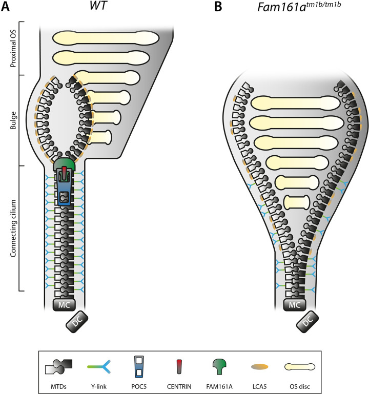
(A) Schematic representation of a part of a WT rod photoreceptor consisting of the CC, the bulge region, and the proximal OS, including its membranous stacked discs. The MTDs are built up from the MC, accompanied by the DC. Cohesion of the MTDs in the CC is maintained by the inner scaffold proteins POC5, CENTRIN, and FAM161A, located at the inner wall of the MTDs, comparable with a closed zipper. Please note that these proteins are found all along the CC, in addition to the MC and DC (not shown in this diagram). MTDs in the CC are connected to the membrane by Y-links, associated with CEP290 and SPATA7 localization. LCA5 localizes to the bulge region, where MTDs are more dispersed due to the absence of the inner scaffold and Y-links. (B) Deficiency of FAM161A causes loss of the entire zip head (the CC inner scaffold) as also POC5 and Centrin are absent, leading to spreading of the MTDs. This spreading, visualized by an open zipper, eventually causes a collapse of the OS structure. Protein localization at the Y-links level is secondarily affected when FAM161A is depleted, as seen by more dispersed CEP290 localization. Furthermore, FAM161A deficiency results in disorganization of the bulge region, obvious from LCA5 localizing more proximal to the MC. Altogether, the CC inner scaffold forms a structural foundation securing proper disc formation and OS integrity. DC, daughter centriole; CC, connecting cilium; MC, mother centriole; MTD, microtubule doublet; OS, outer segment; WT, wild-type.
Comment on
-
The connecting cilium inner scaffold provides a structural foundation that protects against retinal degeneration.PLoS Biol. 2022 Jun 16;20(6):e3001649. doi: 10.1371/journal.pbio.3001649. eCollection 2022 Jun. PLoS Biol. 2022. PMID: 35709082 Free PMC article.
References
Publication types
MeSH terms
LinkOut - more resources
Full Text Sources
