Risk factors for Fontan-associated hepatocellular carcinoma
- PMID: 35714161
- PMCID: PMC9205474
- DOI: 10.1371/journal.pone.0270230
Risk factors for Fontan-associated hepatocellular carcinoma
Abstract
Aims: The incidence of hepatocellular carcinoma (HCC) in patients with Fontan-associated liver disease (i.e., FALD-HCC) has increased over time. However, the risk factors for HCC development remain unclear. Here, we compared the levels of non-invasive markers to the survival rate of FALD-HCC patients.
Methods: From 2003 to 2021, 154 patients (66 men, 42.9%) developed liver disease after undergoing Fontan procedures. HCC was diagnosed in 15 (9.7%) (8 men, 53.3%) at a median age of 34 years (range, 21-45 years). We compared FALD-HCC and non-HCC cases; we generated marker level cutoffs using receiver operating characteristic curves. We sought to identify risk factors for HCC and mortality.
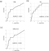
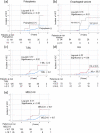
Results: The incidence of HCC was 4.9% in FALD patients within 20 years after the Fontan procedure. Compared with non-HCC patients, FALD-HCC patients exhibited higher incidences of polysplenia and esophageal varices. At the time of HCC development, the hyaluronic acid (HA) level (p = 0.04) and the fibrosis-4 index (p = 0.02) were significantly higher in FALD-HCC patients than in non-HCC patients; the total bilirubin (T-BIL) level (p = 0.07) and the model for end-stage liver disease score [excluding the international normalized ratio (MELD-XI)] (p = 0.06) tended to be higher in FALD-HCC patients. Within approximately 20 years of the Fontan procedure, 10 patients died (survival rate, 96.9%). Kaplan-Meier curve analysis indicated that patients with T-BIL levels ≥ 2.2 mg/dL, HA levels ≥ 55.5 ng/mL, and MELD-XI scores ≥ 18.7 were at high risk of HCC, a generally poor prognosis, and both polysplenia and esophageal varices. Multivariate Cox regression analyses indicated that the complication of polysplenia [Hazard ratio (HR): 10.915] and a higher MELD-XI score (HR: 1.148, both p < 0.01) were independent risk factors for FALD-HCC.
Conclusions: The complication of polysplenia and a MELD-XI score may predict HCC development and mortality in FALD patients.
Conflict of interest statement
KT has received research funding from Sumitomo Dainippon Pharma Co., Ltd., Astellas Pharma Inc., Eisai Co., Ltd., Taiho Pharmaceutical Co., Ltd., Chugai Pharmaceutical Co., Ltd., Daiichi Sankyo Pharmaceutical Co., Ltd., AbbVie GK, Takeda Pharmaceutical Co. Ltd., Asahi Kasei Corporation. Ajinomoto Co., Inc., and Otsuka Pharmaceutical Co., Ltd. The authors received no specific support for this work from a commercial source. We have no declarations relating to employment, consultancy, patents, products in development, marketed products, etc.
Figures


References
-
- Khairy P VG. Late Complications Following the Fontan Operation Diagnosis and Management of Adult Congenital Heart Disease (Third Edition), 2018.
-
- Simonetto DA, Yang HY, Yin M, de Assuncao TM, Kwon JH, Hilscher M, et al.. Chronic passive venous congestion drives hepatic fibrogenesis via sinusoidal thrombosis and mechanical forces. Hepatology. 2015;61(2):648–59. Epub 2015/01/05. doi: 10.1002/hep.27387 ; PubMed Central PMCID: PMC4303520. - DOI - PMC - PubMed
MeSH terms
Substances
Associated data
LinkOut - more resources
Full Text Sources
Medical

