. 2022 Jun 17;18(6):e1010633.
doi: 10.1371/journal.ppat.1010633. eCollection 2022 Jun.

Host cell-dependent late entry step as determinant of hepatitis B virus infection

Affiliations

Host cell-dependent late entry step as determinant of hepatitis B virus infection

Xupeng Hong et al. PLoS Pathog. .

Abstract

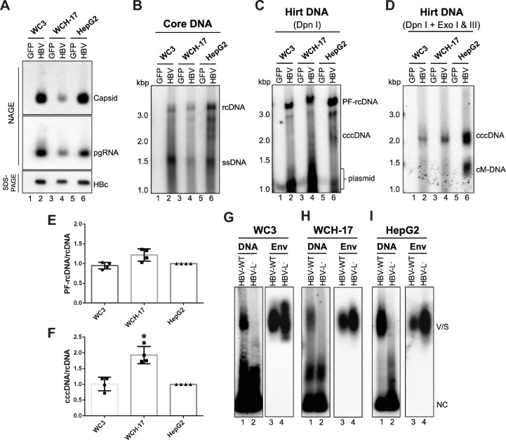
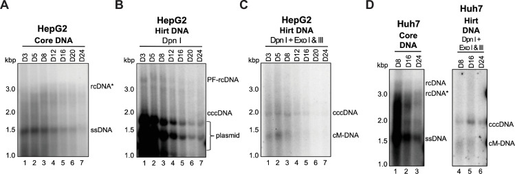
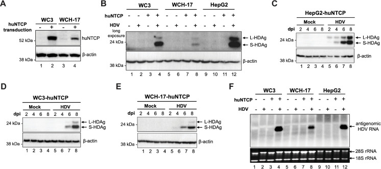
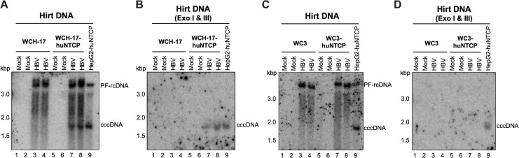
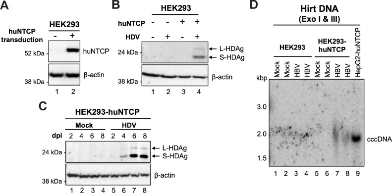
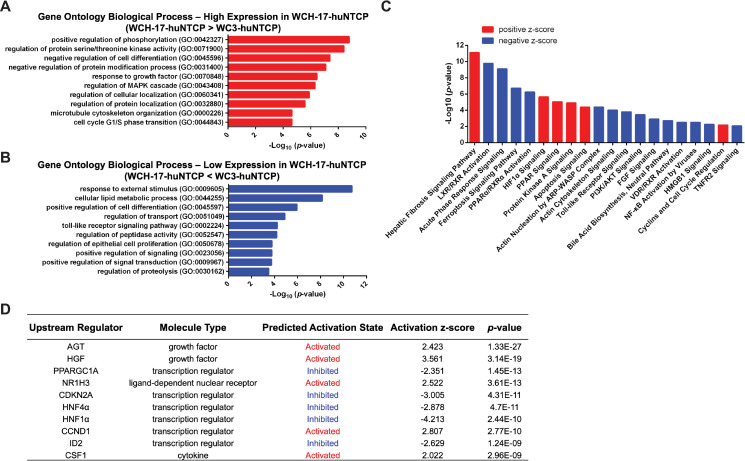
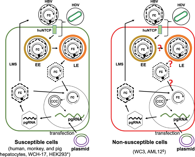
Hepatitis B virus (HBV) has a highly restricted host range and cell tropism. Other than the human sodium taurocholate cotransporting polypeptide (huNTCP), the HBV entry receptor, host determinants of HBV susceptibility are poorly understood. Woodchucks are naturally infected with woodchuck hepatitis virus (WHV), closely related to HBV, but not with HBV. Here, we investigated the capabilities of woodchuck hepatic and human non-hepatic cell lines to support HBV infection. DNA transfection assays indicated that all cells tested supported both HBV and WHV replication steps post entry, including the viral covalently closed circular DNA (cccDNA) formation, which is essential for establishing and sustaining infection. Ectopic expression of huNTCP rendered one, but not the other, woodchuck hepatic cell line and the non-hepatic human cell line competent to support productive HBV entry, defined here by cccDNA formation during de novo infection. All huNTCP-expressing cell lines tested became susceptible to infection with hepatitis D virus (HDV) that shares the same entry receptor and initial steps of entry with HBV, suggesting that a late entry/trafficking step(s) of HBV infection was defective in one of the two woodchuck cell lines. In addition, the non-susceptible woodchuck hepatic cell line became susceptible to HBV after fusion with human hepatic cells, suggesting the lack of a host cell-dependent factor(s) in these cells. Comparative transcriptomic analysis of the two woodchuck cell lines revealed widespread differences in gene expression in multiple biological processes that may contribute to HBV infection. In conclusion, other than huNTCP, neither human- nor hepatocyte-specific factors are essential for productive HBV entry. Furthermore, a late trafficking step(s) during HBV infection, following the shared entry steps with HDV and before cccDNA formation, is subject to host cell regulation and thus, a host determinant of HBV infection.


Conflict of interest statement

The authors have declared that no competing interests exists.

Figures

Fig 1
Fig 1. Woodchuck hepatic cell lines supported the HBV life cycle except entry.
Woodchuck hepatic (hepatoma) cell lines WC3 and WCH-17 and human hepatoma cell line HepG2 were transfected with the HBV replicon (pCIΔA-HBV-HBc) or control (GFP) plasmids, and cells were harvested at 5 days post-transfection. (A) Cytoplasmic lysates were resolved by native agarose gel electrophoresis (NAGE) for comparing capsid assembly (top) and pgRNA packaging (middle) or by SDS-PAGE for comparing HBc expression (bottom). (B) Viral DNA inside nucleocapsids (i.e., core DNA) were released and detected by Southern blot analysis. One twentieth of total core DNA from transfected WC3 and WCH-17 cells and one thirtieth of total core DNA from the transfected HepG2 cells was loaded. PF DNA (i.e., Hirt DNA) was extracted from transfected cells by Hirt extraction method followed by Dpn I treatment to digest input plasmid (C) or with Dpn I and exonuclease I/III (Exo I/III) treatment to remove all DNA except closed circular DNA (D) before agarose gel electrophoresis and Southern blot analysis. One seventh of total WC3 Hirt DNA, one fourth of total WCH-17 Hirt DNA, and one tenth of total HepG2 Hirt DNA extracted from transfected cells was loaded. (E, F) PF-rcDNA and cccDNA signals from each cell line were normalized to the core rcDNA to compare the efficiencies on PF-rcDNA and cccDNA formation (means ± SD, n = 4). Statistical analysis was performed using student’s t test. *, p < 0.05. (G)-(I) Concentrated culture supernatant from transfected WC3 (G), WCH-17 (H), and HepG2 (I) cells were analyzed for virion and HBsAg subviral particle secretion by NAGE and detected by HBV DNA probe, followed by immunoblotting using an anti-HBs polyclonal antibody. ssDNA, single-strand DNA; cM-DNA, closed minus strand circular DNA; Env, envelope protein; V/S, virions and HBsAg subviral particles; NC, nucleocapsids.
Fig 2
Fig 2. Human hepatic cell lines supported WHV cccDNA formation after transfection.
Human hepatoma HepG2 cells were transfected with a WHV replicon, and cells were harvested at the indicated time points for analyzing core DNA (A) and Hirt DNA with Dpn I treatment (B) or with Dpn I plus Exo I/III treatment (C). The same experiment was repeated in another human hepatoma cell line Huh7 (D). ssDNA, single-strand DNA; rcDNA*, rcDNA with incomplete minus strand; cM-DNA, closed minus strand circular DNA.
Fig 3
Fig 3. huNTCP expression in woodchuck hepatic cells conferred susceptibility to HDV infection.
(A) Western blot analysis of deglycosylated cell lysates from parental and huNTCP-expressing cells for validating huNTCP expression. β-actin was used as the loading control. (B) Western blot analysis of whole cell lysates of parental and huNTCP-expressing cells at 8 days post-infection with HDV (ca. 200 genome equivalent per cell) for detecting HDAg using human HDAg antibody. Whole cell lysates of mock-infected or HDV-infected HepG2-huNTCP (C), WC3-huNTCP (D), and WCH-17-huNTCP (E) cells were harvested at 2-, 4-, 6-, and 8-days post-infection and resolved by SDS-PAGE and immunoblotted with human HDAg antibody. (F) Total RNA from mock- or HDV-infected parental or huNTCP-expressing cells were extracted at 8-days post-infection, and 5 μg of total RNA was analyzed by Northern blot for HDV antigenomic RNA. L-HDAg, large HDAg; S-HDAg, small HDAg.
Fig 4
Fig 4. WCH-17 cells were rendered susceptible to HBV infection after huNTCP expression.
WCH-17 (A, B) or WC3 (C, D) parental and huNTCP-expressing cells were plated on regular culture dishes (i.e., with no collagen coating) and infected with ca. 400 genome equivalent of HBV per cell. Three days post infection, the PF DNA (i.e., Hirt DNA) from mock- or HBV-infected cells was extracted by Hirt extraction and treated with Exo I/III followed by Southern blot analysis. Hirt DNA from HBV-infected HepG2-huNTCP cells, loaded at 4-fold less than the Hirt DNA from woodchuck cells, served as the positive control for cccDNA detection.
Fig 5
Fig 5. HEK293 cells were rendered susceptible to HBV and HDV infections after huNTCP expression.
(A) Western blot analysis of deglycosylated cell lysates from parental and huNTCP-expressing HEK293 cells for validating huNTCP expression. β-actin was used as the loading control. (B) SDS-PAGE and western blot analysis of whole cell lysates of parental and huNTCP-expressing cells at 8 days post-infection with HDV for detecting HDAg using the human HDAg antibody. (C) SDS-PAGE and western blot analysis of whole cell lysates of mock-infected or HDV-infected huNTCP-expressing cells were harvested at 2-, 4-, 6-, and 8-days post-infection with the human HDAg antibody. (D) Parental and huNTCP-expressing HEK293 cells were plated on regular culture dishes (i.e., with no collagen coating) and infected with ca. 400 genome equivalent of HBV per cell. Three days post infection, the PF DNA (i.e., Hirt DNA) from mock- or HBV-infected cells was extracted by Hirt extraction and treated with Exo I/III followed by Southern blot analysis. Hirt DNA from HBV-infected HepG2-huNTCP cells, loaded at 4-fold less than the Hirt DNA from HEK293 and HEK-293-huNTCP cells, served as the positive control for cccDNA detection.
Fig 6
Fig 6. Comparative transcriptomic analysis of WC3 and WCH-17 huNTCP-expressing cells.
(A) Gene Ontology (GO) analysis of biological processes for genes expressed at higher levels in WCH-17-huNTCP cells. (B) GO analysis of biological processes for genes expressed at lower levels in WCH-17-huNTCP cells. (C) The most highly regulated canonical pathways by IPA analysis of genes differentially expressed in WCH-17-huNTCP cells relative to WC3-huNTCP cells. Negative z-score (blue) or positive z-score (red) indicate the pathways that were likely inhibited or activated in WCH-17-huNTCP cells compared to WC3-huNTCP cells, respectively. (D) The top 10 upstream regulators and their predicted activation status in WCH-17-huNTCP cells (relative to WC3-huNTCP cells) in IPA analysis of transcriptomic results.
Fig 7
Fig 7. Model of HBV infection in susceptible and non-susceptible cells.
WCH-17 and HEK293 (left) and WC3 (right) cells could support HBV cccDNA formation via the intracellular amplification pathway after HBV replicon transfection. Ectopic expression of huNTCP in WCH-17 and HEK293 cells (and other cells, see text) allowed HBV infection as evidenced by the establishment of cccDNA in the nucleus of infected cells. However, huNTCP expression in WC3 cells failed to support HBV infection, indicating an infection block at a step(s) after initial entry steps shared with HDV (in bracket) and prior to the nuclear import of rcDNA, the common step for cccDNA formation shared between infection and intracellular amplification. rc, relaxed circular DNA; ccc, covalently closed circular DNA; EE, early endosome; LE, late endosome; LMS, larger, middle, and small surface proteins. *, HEK293 cells cannot support HBV gene expression due to the lack of liver-specific transcription factors. $, AML12 cells support efficient HBV cccDNA formation via intracellular amplification but are only weakly susceptible to HBV infection after huNTCP expression.?, potential steps during HBV infection that may be blocked in WC3 cells. See text for details.

Similar articles

Cited by

References

    1. WHO. Hepatitis B: World Health Organization; 2021. Available from: https://www.who.int/news-room/fact-sheets/detail/hepatitis-b.
    1. Hu J, Protzer U, Siddiqui A. Revisiting Hepatitis B Virus: Challenges of Curative Therapies. Journal of virology. 2019;93(20):e01032–19. PubMed Central PMCID: PMC6798116. doi: 10.1128/JVI.01032-19 - DOI - PMC - PubMed
    1. Hu J, Lin Y-Y, Chen P-J, Watashi K, Wakita T. Cell and animal models for studying hepatitis B virus infection and drug development. Gastroenterology. 2019;156(2):338–54. PubMed Central PMCID: PMC6649672. doi: 10.1053/j.gastro.2018.06.093 - DOI - PMC - PubMed
    1. Burwitz BJ, Zhou Z, Li W. Animal models for the study of human hepatitis B and D virus infection: new insights and progress. Antiviral Research. 2020:104898. doi: 10.1016/j.antiviral.2020.104898 - DOI - PubMed
    1. Hu J, Seeger C. Hepadnavirus Genome Replication and Persistence. Cold Spring Harb Perspect Med. 2015;5(7):a021386. Epub 2015/07/03. doi: 10.1101/cshperspect.a021386 ; PubMed Central PMCID: PMC4484952. - DOI - PMC - PubMed

Publication types