Review
. 2022 Jun 16;82(12):2236-2251.
doi: 10.1016/j.molcel.2022.05.029.

Hidden codes in mRNA: Control of gene expression by m6A

Affiliations
Review

Hidden codes in mRNA: Control of gene expression by m6A

Shino Murakami et al. Mol Cell. .

Abstract

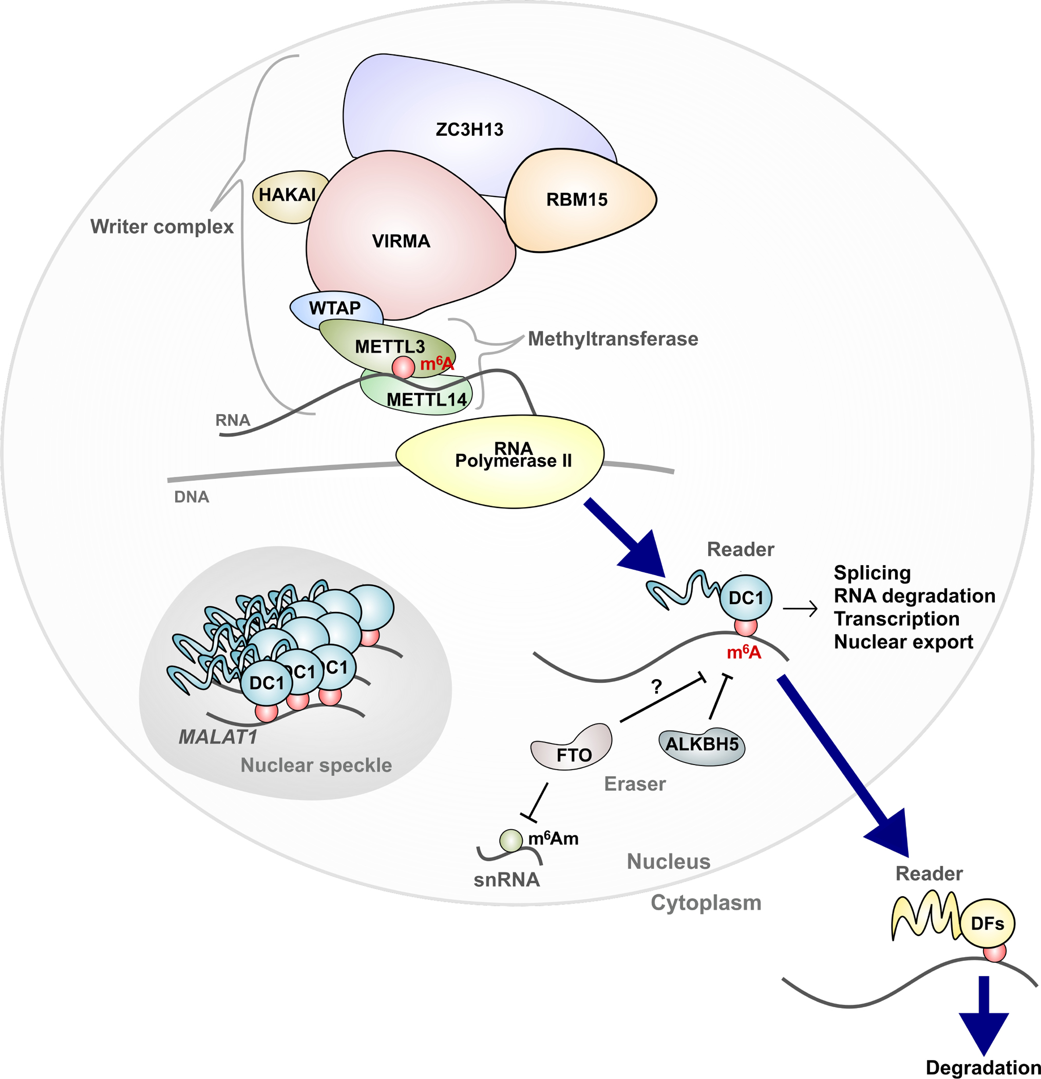
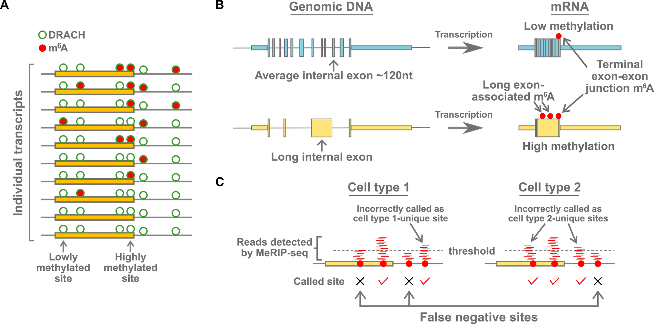
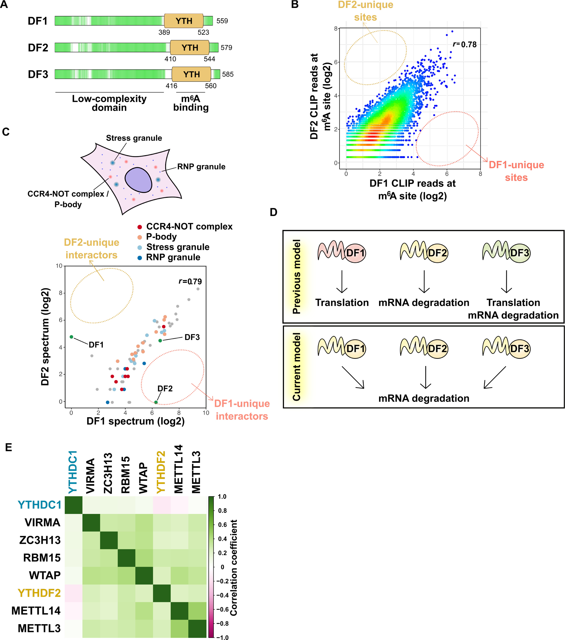
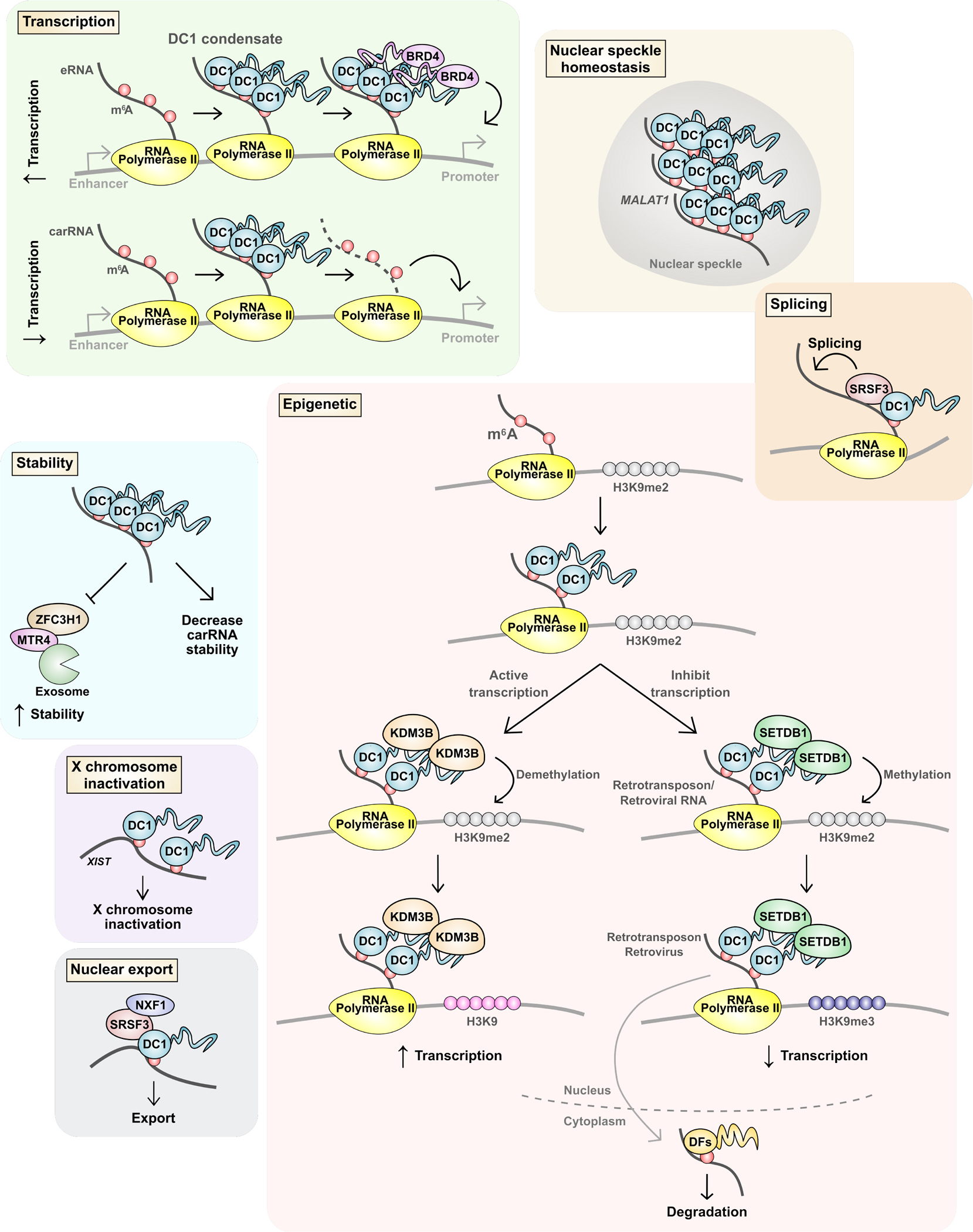
Information in mRNA has largely been thought to be confined to its nucleotide sequence. However, the advent of mapping techniques to detect modified nucleotides has revealed that mRNA contains additional information in the form of chemical modifications. The most abundant modified nucleotide is N6-methyladenosine (m6A), a methyl modification of adenosine. Although early studies viewed m6A as a dynamic and tissue-specific modification, it is now clear that the mRNAs that contain m6A and the location of m6A in those transcripts are largely universal and are influenced by gene architecture, i.e., the size and location of exons and introns. m6A can affect nuclear processes such as splicing and epigenetic regulation, but the major effect of m6A on mRNAs is to promote degradation in the cytoplasm. m6A marks a functionally related cohort of mRNAs linked to certain biological processes, including cell differentiation and cell fate determination. m6A is also enriched in other cohorts of mRNAs and can therefore affect their respective cellular processes and pathways. Future work will focus on understanding how the m6A pathway is regulated to achieve control of m6A-containing mRNAs.

Keywords: METTL14; METTL3; N6-methyladenosine; RBM15; RNA degradation; RNA stability; VIRMA; WTAP; YTHDC1; YTHDC2; YTHDF1; YTHDF2; YTHDF3; ZC3H13; epitranscriptome; m(6)A.


Conflict of interest statement

Declaration of interests S.R.J. is an advisor to, and owns equity in, 858 Therapeutics. S.R.J. is an author of patents related to detection and use of m(6)A in mRNA.

Figures

Figure 1.
Figure 1.. Writing and reading m6A
m6A is deposited co-transcriptionally by a writer complex. The catalytic subunit METTL3 forms a heterodimer with METTL14 to form the minimal methyltransferase core complex. METTL3/14 is a component of a larger writer complex, and each protein is required for m6A deposition in the cell. YTHDC1 (DC1), a nuclear m6A-binding protein, is assembled into nuclear condensates via m6A-containing RNA, including the nuclear noncoding RNA MALAT1. These condensates, such as nuclear speckles, are important for various processing steps that most mRNAs undergo in the nucleus. DC1 also binds and regulates specific m6A RNA to regulate splicing in some transcripts, or to influence epigenetic marks, transcription, RNA stability, or nuclear export. In the nucleus, m6A may be erased, particularly in testes where the nuclear m6A eraser ALKBH5 is enriched. FTO is an efficient demethylase for m6Am in snRNA, and may also demethylate m6A. Once exported to the cytoplasm, m6A is bound by DF proteins, which comprise YTHDF1, YTHDF2, and YTHDF3, which all promote mRNA degradation (Bawankar et al., 2021; Schöller et al., 2018; Knuckles et al., 2018).
Figure 2.
Figure 2.. Deposition and detection of m6A
(A) m6A is a low stoichiometry modification. At any given m6A site in a specific mRNA, only a small number (usually less than 20%) of the transcript copies in the cell will have m6A at that site. Most of the DRACH m6A consensus sites are not methylated (green circle). Additionally, some DRACH sites are more methylated than others, and all DRACH sites are likely methylated to some degree, although the stoichiometry is likely very low. The molecular mechanism that causes some DRACH sites to be more methylated than others, even in the same transcript, are not fully understood. Although m6A mapping may reveal multiple m6A sites for a given mRNA, the individual transcripts that comprise the m6A annotation are typically methylated only at a subset of sites. Newer single-transcript analysis methods have revealed the stochastic nature of methylation on individual transcripts. (B) Gene architecture influences m6A deposition. Shown (left) are examples of two genes, one containing a long internal exon. After transcription (right), m6A is preferentially formed on the regions of transcripts corresponding to long exons. m6A is also enriched near the terminal exon-exon junction, particularly in mRNAs with long 3’UTRs. The correlation of m6A with these genomic features suggests that the writer complex is regulated by other events that are responsive to gene architecture. (C) Putative differences in m6A are sometimes artifacts of the methods used to call m6A sites from mapping data. Many studies have used mapping methods such as MeRIP-seq to map m6A and to compare transcriptome-wide distributions of m6A between two different cell conditions. These methods map reads (red lines) immunoprecipitated by an m6A-binding antibody. m6A sites are often called based on whether there are a sufficient number of reads above a threshold. However, the number of reads that map to any m6A sites can be very variable, even between replicates (McIntyre et al., 2020). Thus, the same m6A site in one sample might be called in one sample, but not the other, if the number of reads just passes or misses the threshold. For this reason, the differences in the transcriptome-wide distribution of m6A in many experiments may have been highly overestimated.
Figure 3.
Figure 3.. YTHDF proteins function together to promote degradation of m6A-marked mRNAs
(A) DF proteins are low-complexity domain-containing “m6A reader” proteins. All three paralogs are highly similar and contain a proline-, glutamine-, and asparagine-rich low-complexity domain (green) followed by m6A-binding YTH domain. The low-complexity domain allows these three DF proteins to localize to granule structures such as RNP granules, P-body, and stress granules. (B) All DF paralogs bind to all m6A sites, with no DF paralog showing preferential binding to any m6A site. Shown is a pairwise comparison of DF1 and DF2 iCLIP coverage at each single nucleotide-resolved m6A site in HEK293T cells. The color indicates the density. r, Pearson correlation coefficient. Similar results are seen when comparing DF3 to either DF1 or DF2 (Zaccara and Jaffrey, 2020). (C) All three DF paralogs likely serve the same function since they interact with same sets of proteins. Pairwise comparison of the probabilities of interaction for DF1- and DF2-interacting proteins determined by proximal labeling and proteomics (Youn et al., 2018). Proximal proteins were detected by DF1 or DF2 C-terminally tagged with promiscuous biotin ligase (BirA). The average probability of interaction for either DF1 or DF2 or both higher than 0.9 are shown. r, Pearson correlation coefficient. (D) Revised model of the YTHDF protein function. DF1 and DF3 were previously shown to promote translation, while DF2 and DF3 enhance mRNA degradation. More recent studies show all three DF proteins redundantly promote mRNA degradation. (E) DF2 is the major protein mediating the effects of m6A on cell survival and proliferation. DepMap analysis reveals genes that show similar patterns of gene dependency as METTL3 across over 1000 different cell types. Members of the writer complex show the highest similarity to METTL3 in the different cell lines. The DF2 also shows a similar type of gene dependency as METTL3. In contrast, the pattern of cell dependency on DC1 shows poor correlation with METTL3. Dependency scores for m6A writers and readers for each cell line were analyzed by pairwise comparison. Pearson correlation coefficients were visualized in the matrix heatmap upon hierarchical clustering.
Figure 4.
Figure 4.. DC1 regulates diverse nuclear processing events through m6A
DC1 has been reported to exert a wide range of effects on mRNA. Some studies suggest transcription regulation by DC1. DC1 can enhance transcription by binding m6A-containing enhancer RNAs (eRNAs). DC1 forms a condensate with eRNAs that recruits BRD4. DC1 has also been shown to bind “chromatin-associated regulatory RNAs” (carRNAs), which broadly refers to many types of nuclear RNAs including eRNAs. DC1 was shown to mediate the degradation of carRNAs and subsequently reduce nearby gene expression. DC1 also has been shown to increase and decrease methylation of H3K9. In some studies, DC1 binds m6A mRNA and recruits KDM3B to demethylate H3K9me2 and enhance transcription. Other studies show that DC1 binds retrotransposon RNA and recruits SETDB1 to form H3K9me3 to reduce transcription. DC1 has also been shown to control splicing, RNA stability, nuclear export, and to promote X chromosome inactivation. DC1 also binds to the non-coding RNA MALAT1 which allows DC1 to have an important role in maintaining the protein composition of nuclear speckles.

References

    1. Agarwala SD, Blitzblau HG, Hochwagen A, and Fink GR (2012). RNA methylation by the MIS complex regulates a cell fate decision in yeast. PLoS Genet 8, e1002732. 10.1371/journal.pgen.1002732 - DOI - PMC - PubMed
    1. PGENETICS-D-12–00168 [pii].
    1. Bartosovic M, Molares HC, Gregorova P, Hrossova D, Kudla G, and Vanacova S (2017). N6-methyladenosine demethylase FTO targets pre-mRNAs and regulates alternative splicing and 3’-end processing. Nucleic Acids Res 45, 11356–11370. 10.1093/nar/gkx778. - DOI - PMC - PubMed
    1. Batista PJ, Molinie B, Wang J, Qu K, Zhang J, Li L, Bouley DM, Lujan E, Haddad B, Daneshvar K, et al. (2014). m(6)A RNA modification controls cell fate transition in mammalian embryonic stem cells. Cell Stem Cell 15, 707–719. 10.1016/j.stem.2014.09.019. - DOI - PMC - PubMed
    1. Bawankar P, Lence T, Paolantoni C, Haussmann IU, Kazlauskiene M, Jacob D, Heidelberger JB, Richter FM, Nallasivan MP, Morin V, et al. (2021). Hakai is required for stabilization of core components of the m6A mRNA methylation machinery. Nat Commun 12, 3778. 10.1038/s41467-021-23892-5. - DOI - PMC - PubMed

Publication types

LinkOut - more resources