Review
. 2022 Jun 16;82(12):2298-2314.
doi: 10.1016/j.molcel.2022.05.004.

Hallmarks of DNA replication stress

Affiliations
Review

Hallmarks of DNA replication stress

Sneha Saxena et al. Mol Cell. .

Abstract

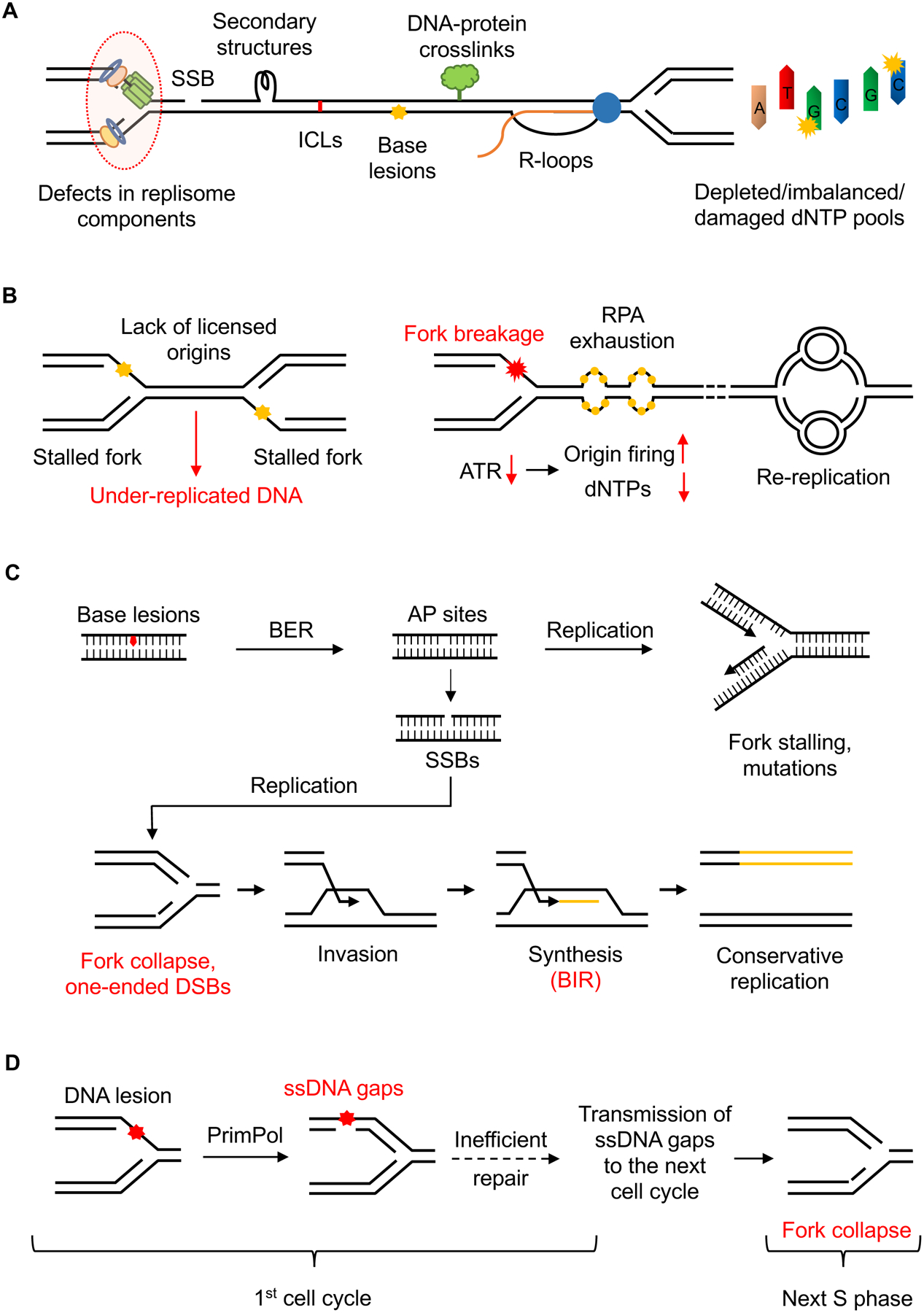
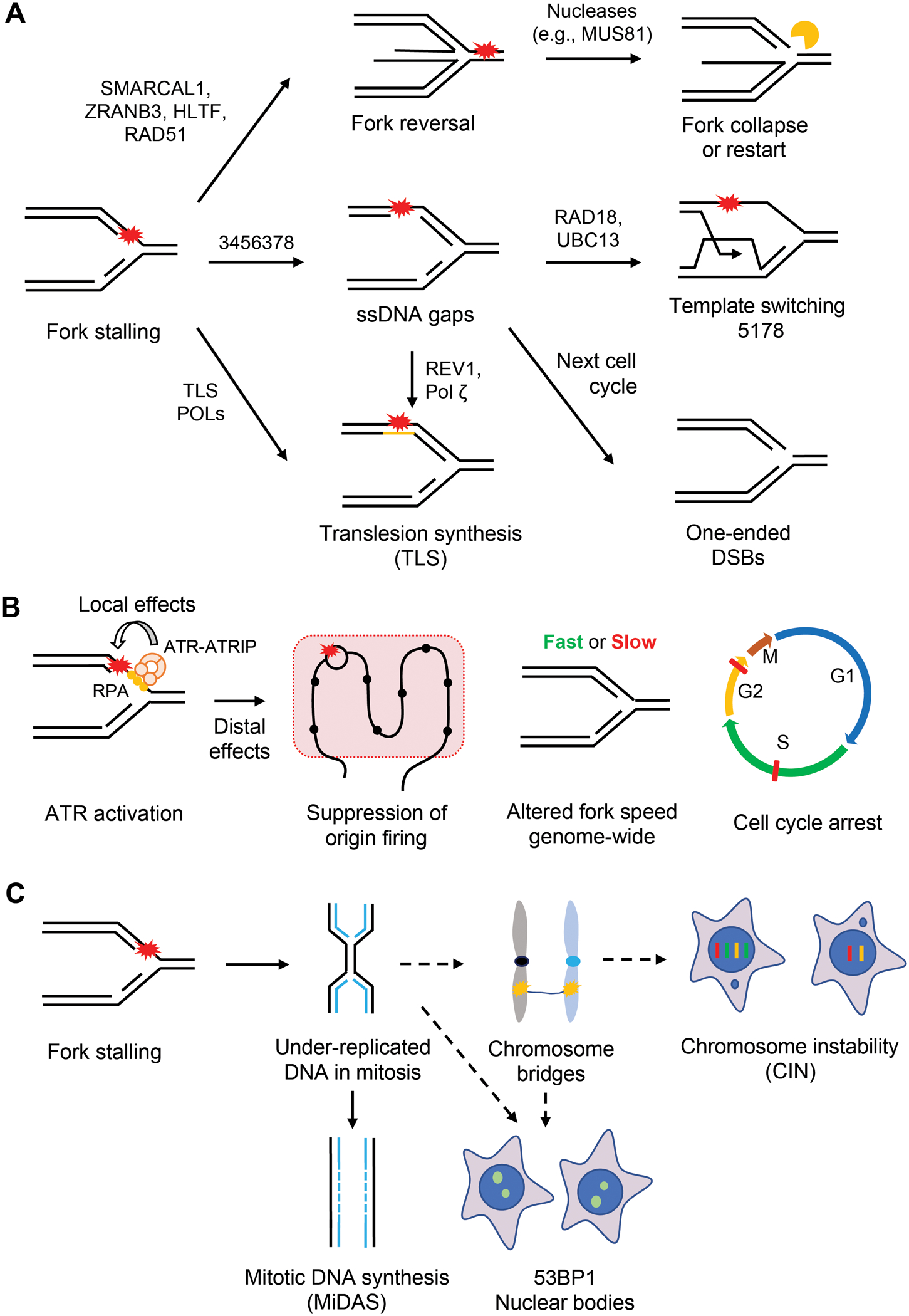
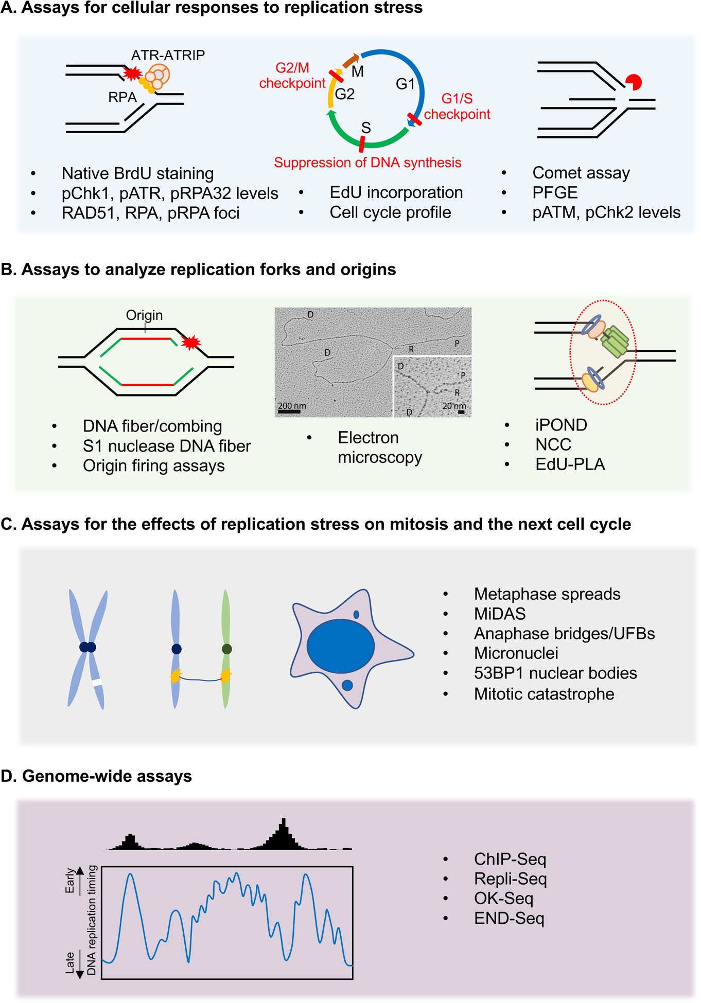
Faithful DNA replication is critical for the maintenance of genomic integrity. Although DNA replication machinery is highly accurate, the process of DNA replication is constantly challenged by DNA damage and other intrinsic and extrinsic stresses throughout the genome. A variety of cellular stresses interfering with DNA replication, which are collectively termed replication stress, pose a threat to genomic stability in both normal and cancer cells. To cope with replication stress and maintain genomic stability, cells have evolved a complex network of cellular responses to alleviate and tolerate replication problems. This review will focus on the major sources of replication stress, the impacts of replication stress in cells, and the assays to detect replication stress, offering an overview of the hallmarks of DNA replication stress.

Keywords: ATR; DNA damage; DNA repair; DNA replication; cancer; cell cycle; checkpoint; genomic integrity; genomic stability; replication stress.


Conflict of interest statement

Declaration of interests The authors declare no competing interests. L.Z. is a member of the advisory board of Molecular Cell.

Figures

Figure 1.
Figure 1.. Sources of DNA replication stress.
(A) The progression of replication forks can be impeded by several obstacles including SSBs, DNA secondary structures, ICLs, etc. Defects in replisome components or alterations in dNTP pools can also reduce fork speed, compromise fork stability, and promote mutagenesis. (B) Lack of licensed origins leads to reduced rescue of stalled forks, resulting in under-replicated regions. Increased origin firing reduces fork speed by depleting dNTPs and other replisome factors. It also generates excessive amount of ssDNA, which exhausts the nuclear pool of RPA and induces genome-wide breakage of replication forks. Re-replication forks progress slowly and undergo “head-to-tail” collisions with previously initiated replication forks, resulting in DSBs. (C) Excessive repair intermediates like AP sites and SSBs act as roadblocks to replication forks. Collisions of active forks with SSBs cause fork collapse and one-ended DSBs, which can be recovered by BIR, an aberrant replication process that is error-prone and generates replication stress. (D) Replication forks can bypass DNA lesions by PrimPol-mediated repriming, which generates ssDNA gaps behind the forks. If unrepaired, these ssDNA gaps can persist into the next cell cycle, where they encounter active forks and cause fork collapse, generating trans-cell cycle replication stress.
Figure 2.
Figure 2.. Consequences of DNA replication stress.
(A) Obstructions to replication progression cause fork stalling. Several replication-coupled repair mechanisms are used to promote the bypass and/or repair of DNA damage. Stalled forks can be reversed by the action of DNA translocases such as ZRANB3, HLTF, and SMARCAL1 and recombinase RAD51. The resulting four-way structure can be cleaved by nucleases like MUS81 to form DSBs. The DSBs at reversed forks can promote fork restart, but can also be toxic when they accumulate at high levels. Alternatively, stalled forks can bypass DNA lesions by PrimPol-mediated repriming. Repriming generates ssDNA gaps behind the fork, which can be repaired by TS or TLS in S and G2 phases. In the event of inefficient repair, these gaps persist into the next S phase and induce one-ended DSBs. Stalled forks can also directly bypass DNA lesions through TLS. (B) Fork stalling generates stretches of ssDNA at stressed forks, which is coated by the RPA complex. ssDNA-RPA in turn recruits the ATR-ATRIP kinase complex to stalled forks. Once activated, ATR phosphorylates substrates at stalled forks to promote fork stabilization and restart (local effects). Furthermore, ATR suppresses origin firing, alters fork speed genome-wide, and induces cell cycle arrests in S and G2/M phases (distal effects). (C) Under-replicated DNA (UR-DNA) can persist into mitosis and undergo MiDAS. If not replicated by MiDAS, UR-DNA forms chromosome or DNA bridges in anaphase. If these bridges are not resolved by repair proteins, they give rise to chromosome instability (CIN). If UR-DNA is segregated into daughter cells, it is sequestered into 53BP1 nuclear bodies (53BP1 NBs).
Figure 3.
Figure 3.. Assays for the detection of replication stress.
(A) Cellular assays to study the global effects of replication stress by measuring the accumulation of ssDNA, phosphorylation of ATR substrates, suppression of DNA synthesis, cell cycle arrests, and generation of DSBs. (B) DNA fiber-based assays directly study the impact of replication stress on progressing forks and origin firing. Replication forks can be directly visualized by EM to study fork reversal and ssDNA gap generation. The EM image is reproduced from (Genois et al. 2021). Other assays including iPOND and PLA are used to analyze factors associated with nascent DNA. (C) Cellular assays that use mitotic defects as a proxy to measure the replication stress in the preceding S phase. These assays study the effects of replication stress on mitosis and the next cell cycle in the form of anaphase bridges, micronucleation, chromosomal aberrations, and 53BP1 NBs. (D) Genomic assays that use next-generation sequencing (NGS) to obtain a whole-genome view of the effects of replication stress. Theses assays are used to map the regions of replication perturbation across the genome and to understand the contributions of specific chromatin environments and DNA sequence elements to replication stress.

Similar articles

Cited by

References

    1. Abe T, Kawasumi R, Giannattasio M, Dusi S, Yoshimoto Y, Miyata K, Umemura K, Hirota K, and Branzei D (2018). AND-1 fork protection function prevents fork resection and is essential for proliferation. Nat Commun 9, 3091. - PMC - PubMed
    1. Aguilera A, and Garcia-Muse T (2012). R loops: from transcription byproducts to threats to genome stability. Mol Cell 46, 115–124. - PubMed
    1. Ait Saada A, Teixeira-Silva A, Iraqui I, Costes A, Hardy J, Paoletti G, Freon K, and Lambert SAE (2017). Unprotected Replication Forks Are Converted into Mitotic Sister Chromatid Bridges. Mol Cell 66, 398–410 e394. - PubMed
    1. Alabert C, Bukowski-Wills JC, Lee SB, Kustatscher G, Nakamura K, de Lima Alves F, Menard P, Mejlvang J, Rappsilber J, and Groth A (2014). Nascent chromatin capture proteomics determines chromatin dynamics during DNA replication and identifies unknown fork components. Nat Cell Biol 16, 281–293. - PMC - PubMed
    1. Almeida R, Fernandez-Justel JM, Santa-Maria C, Cadoret JC, Cano-Aroca L, Lombrana R, Herranz G, Agresti A, and Gomez M (2018). Chromatin conformation regulates the coordination between DNA replication and transcription. Nat Commun 9, 1590. - PMC - PubMed

Publication types

Substances

LinkOut - more resources