. 2022 Jul 12;17(7):1546-1560.
doi: 10.1016/j.stemcr.2022.05.014. Epub 2022 Jun 16.

CD49b identifies functionally and epigenetically distinct subsets of lineage-biased hematopoietic stem cells

Affiliations

CD49b identifies functionally and epigenetically distinct subsets of lineage-biased hematopoietic stem cells

Ece Somuncular et al. Stem Cell Reports. .

Abstract

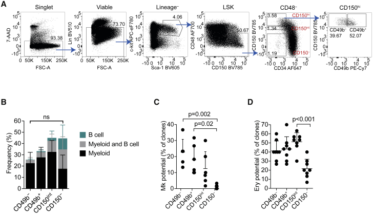
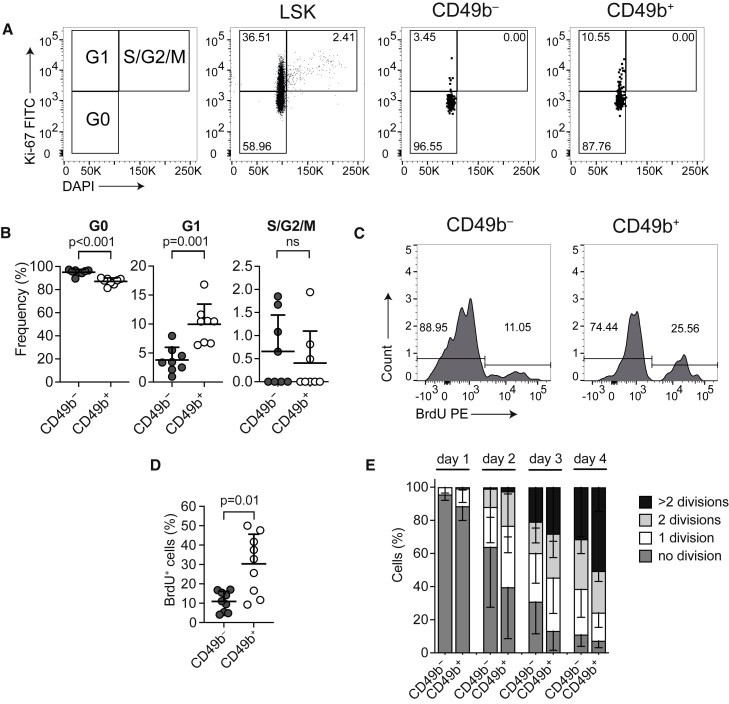
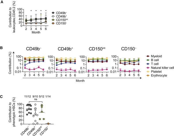
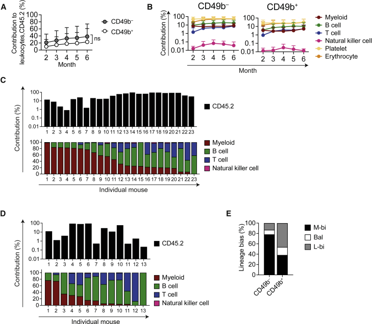
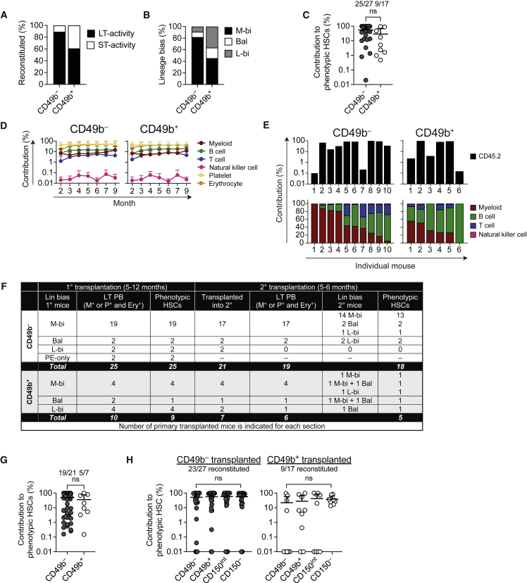
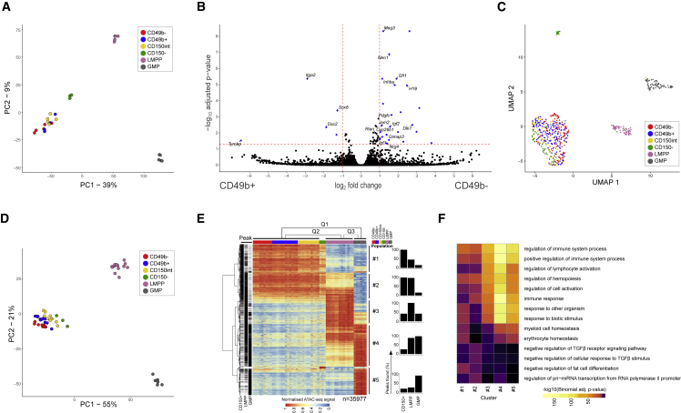
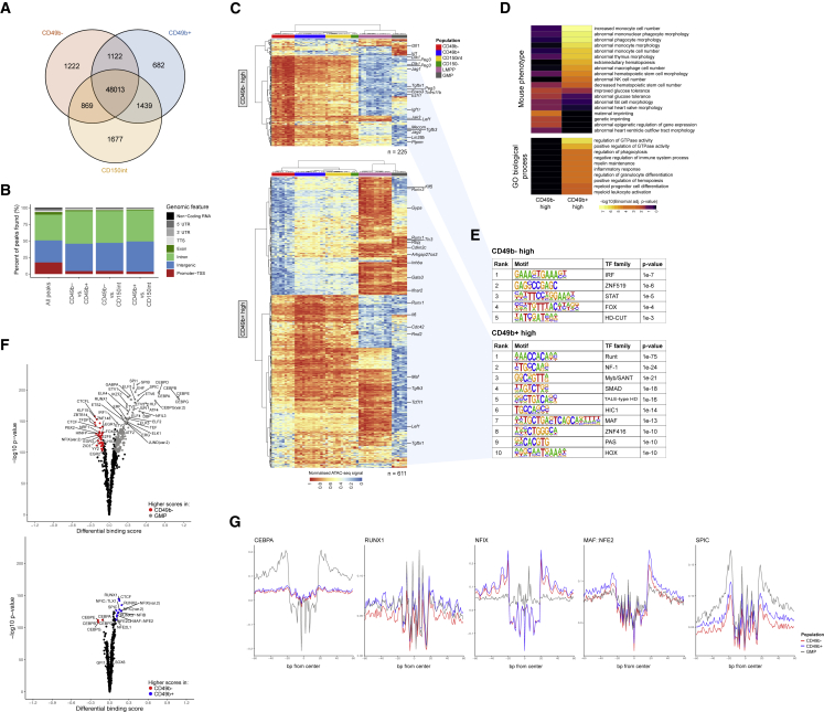
Hematopoiesis is maintained by functionally diverse lineage-biased hematopoietic stem cells (HSCs). The functional significance of HSC heterogeneity and the regulatory mechanisms underlying lineage bias are not well understood. However, absolute purification of HSC subtypes with a pre-determined behavior remains challenging, highlighting the importance of continued efforts toward prospective isolation of homogeneous HSC subsets. In this study, we demonstrate that CD49b subdivides the most primitive HSC compartment into functionally distinct subtypes: CD49b- HSCs are highly enriched for myeloid-biased and the most durable cells, while CD49b+ HSCs are enriched for multipotent cells with lymphoid bias and reduced self-renewal ability. We further demonstrate considerable transcriptional similarities between CD49b- and CD49b+ HSCs but distinct differences in chromatin accessibility. Our studies highlight the diversity of HSC functional behaviors and provide insights into the molecular regulation of HSC heterogeneity through transcriptional and epigenetic mechanisms.

Keywords: ATAC-seq; CD49b; HSC heterogeneity; RNA-seq; epigenetic regulation; hematopoietic stem cells; lineage bias; single-cell transplantation.


Figures

Figure 1
Figure 1
The HSC compartment can be further subfractionated with CD49b (A) Fluorescence-activated cell sorting (FACS) profile and gating strategy of phenotypic HSC subsets and further separation with CD49b in CD117-enriched BM cells. Frequencies of parent gates are shown. (B) In vitro differentiation potential of single sorted cells to myeloid (CD11b+Gr-1+ and/or CD11b+F4/80+) and B cells (B220+CD19+, nCD49b = 568 cells, nCD49b+ = 536 cells, nCD150int = 401 cells, nCD150 = 409 cells; nine replicates; five independent experiments). ns, not significant. (C) Megakaryocyte differentiation culture of single plated cells (n = 360 cells/population, six replicates, three independent experiments). (D) Erythroid colony-forming assay of CD49b, CD49b+, CD150int, and CD150 cells (n = 8 replicates/population, 30 cells per replicate, two independent experiments). Mean ± SD is shown in (B–D). Statistical significance in (B) was calculated based on total cloning frequency. See also Figure S1.
Figure 2
Figure 2
CD49b and CD49b+ subsets have different cell-cycle and cell-proliferation kinetics (A) Cell-cycle analysis of CD117-enriched BM cells. The frequency of G0, G1, and S/G2/M cells in a representative mouse is shown. (B) Frequency of CD49b and CD49b+ HSCs in G0, G1, and S/G2/M (n = 8 mice, two independent experiments). ns, not significant. (C) BrdU analysis of CD117-enriched BM cells. The frequency of BrdU and BrdU+ cells in a representative mouse is shown. (D) Frequency of BrdU+ CD49b and CD49b+ HSCs (n = 9 mice, three independent experiments). (E) Cell divisions from cultured single cells on days 1–4 (nCD49b = 347 cells, nCD49b+ = 370 cells, five replicates, three independent experiments). Mean ± SD is shown in (B), (D), and (E).
Figure 3
Figure 3
CD49b and CD49b+ HSCs are the most durable subsets (A and B) Total donor contribution (A) and donor contribution to platelets, erythrocytes, and myeloid, B, T, and natural killer cells (B) in peripheral blood (PB) from five cell transplantations (nCD49b = 28 mice, nCD49b+ = 22 mice, nCD150int = 12 mice, nCD150 = 13 mice, three independent experiments). (C) Donor contribution to phenotypic HSCs (LSKFlt-3CD48CD150+ or LSKCD48CD150+), 5–6 months after five cell transplantations (nCD49b = 12 mice, nCD49b+ = 10 mice, nCD150int = 12 mice, nCD150 = 13 mice, three independent experiments). The numbers of reconstituted mice out of all mice analyzed are indicated. ns, not significant. Asterisks indicate statistically significant differences: p = 0.028, CD49b versus CD150, and p = 0.05, CD150int versus CD150, month 2; p = 0.032, CD49b versus CD150, and p = 0.006, CD150int versus CD150, month 3; p = 0.015, CD49b versus CD150, and p = 0.005, CD150int versus CD150, month 4; p = 0.005, CD49b versus CD150, and p = 0.013, CD150int versus CD150, month 5; and p = 0.016, CD49b versus CD49b+, month 6. Mean ± SD is shown in (A−C). See also Figure S2.
Figure 4
Figure 4
CD49b and CD49b+ HSCs reconstitute all blood lineages, but at different ratios (A and B) Total donor contribution (A) and donor contribution to platelets, erythrocytes, and myeloid, B, T, and natural killer cells (B) in PB from single-cell transplantation (nCD49b = 28 mice, nCD49b+ = 18 mice, five independent experiments). ns, not significant. (C and D) Total donor contribution (top) and relative contribution to myeloid, B, T, and natural killer cells (bottom) in PB of CD49b (C) or CD49b+ (D) single-cell-transplanted mice, 5–6 months post-transplantation (nCD49b = 23 mice, nCD49b+ = 13 mice, five independent experiments). (E) Lineage bias categorization of single-cell-transplanted mice 5–6 months post-transplantation (nCD49b = 23 mice, nCD49b+ = 13 mice, five independent experiments). M-bi, myeloid-biased; Bal, balanced; L-bi, lymphoid-biased. Mean ± SD is shown (A and B). See also Figures S3–S5.
Figure 5
Figure 5
CD49b HSCs are the most durable stem cells (A) Short-term (ST) or long-term (LT) HSC activity of reconstituted single-cell-transplanted mice (nCD49b = 28 mice, nCD49b+ = 18 mice, five independent experiments). LT defined as myeloid (M)+ or platelet (P)+ and erythrocyte (Ery)+ in PB, 5–6 months post-transplantation. (B) Lineage bias distribution of single-cell-transplanted mice with LT activity in (A) (nCD49b = 25 mice, nCD49b+ = 11 mice, five independent experiments). (C) Donor contribution to phenotypic HSCs (LSKFlt-3CD48CD150+ or LSKCD48CD150+) 5–12 months after single-cell transplantation (nCD49b = 27 mice, nCD49b+ = 17 mice, five independent experiments). ns, not significant. (D) Donor contribution to platelets, erythrocytes, and myeloid, B, T, and natural killer cells in PB from single-cell transplantation (nCD49b = 10 mice, nCD49b+ = 6 mice, two independent experiments). (E) Total donor contribution (top) and relative contribution to myeloid, B, T, and natural killer cells (bottom) in PB of CD49b and CD49b+ single-cell-transplanted mice, 9 months post-transplantation (nCD49b = 10 mice, nCD49b+ = 6 mice, two independent experiments). (F) Overview of CD49b and CD49b+ primary reconstituted single-cell-transplanted mice with LT activity from (A). The categorized lineage bias and number of mice with donor contribution to PB and phenotypic HSCs (LSKFlt-3CD48CD150+ or LSKCD48CD150+) 5–12 months post-transplantation, in primary and secondary transplantation, are indicated. (G) Donor contribution to phenotypic HSCs (LSKFlt-3CD48CD150+ or LSKCD48CD150+) in secondary transplantation with whole BM cells, 5–6 months post-transplantation (nCD49b = 50, nCD49b+ = 8 mice, five independent experiments). ns, not significant. (H) Donor contribution to phenotypic lineage-biased HSC subsets, 5–12 months post-transplantation, from single-cell-transplanted mice (nCD49b = 27 mice, nCD49b+ = 17 mice, five independent experiments). The number of reconstituted primary donor mice out of all mice analyzed is indicated in (C), (G) and (H). Mean ± SD is shown (C, D, G, H). ns, not significant. See also Figures S4 and S5.
Figure 6
Figure 6
The CD49b and CD49b+ subsets are transcriptionally similar but epigenetically distinct (A) PCA of bulk RNA-seq data (nCD49b = 5, nCD49b+ = 4, nCD150int = 5, nCD150 = 3, nLMPP = 5, nGMP = 5 replicates, five independent experiments). (B) Volcano plot of differential expression between CD49b and CD49b+ subsets. Differentially expressed genes (padj < 0.05, |fold change| > 2) are marked in blue, with selected genes annotated. (C) UMAP visualization of single-cell RNA-seq data colored by cell population (nCD49b = 135, nCD49b+ = 146, nCD150int = 59, nCD150 = 77, nLMPP = 57, nGMP = 74 cells, two independent experiments). (D) PCA of ATAC-seq data (nCD49b = 9, nCD49b+ = 12, nCD150int = 10, nCD150 = 3, nLMPP = 13, nGMP = 6 replicates, seven independent experiments). (E) Heatmap (left) of row-normalized chromatin accessibility for differential regions (padj < 1 × 10−4, |fold change| > 2) in pairwise comparisons between CD150+, LMPP, and GMP populations. Regions are divided into five clusters based on hierarchical clustering. Bar graphs (right) show how many peaks were called in each cluster. (F) GO enrichment analysis of clusters 1–5. The top three significantly enriched terms for each cluster from the GO biological process are shown. See also Figures S6 and S7.
Figure 7
Figure 7
The CD49b and CD49b+ subsets exhibit distinct epigenetic changes (A) Overlap of open chromatin regions between CD150+ populations. Peaks with a normalized read count of >1.5 in more than one-third of samples were considered found. (B) Genomic feature distribution of regions with differential accessibility (padj < 0.01) and all regions as a reference ("all peaks"). (C) Heatmap of row-normalized chromatin accessibility for regions with differential accessibility (padj < 0.01) between CD49b (top; n = 225 peaks) and CD49b+ (bottom; n = 611 peaks) subsets. For selected regions the nearest gene is indicated. (D) GO enrichment analysis of regions with differential accessibility between CD49b and CD49b+ subsets. The top 10 significantly enriched terms from the mouse phenotype and GO biological process are shown. (E) Transcription factor (TF) families with enriched binding motifs (q <0.01, top 10) in regions with increased accessibility in CD49b (top) or CD49b+ (bottom) cells. (F) Volcano plots of differential TF binding. Transcription factors with differential binding activity (differential binding score >0.1, p < 1 × 10−100) are colored and selectively annotated. (G) Aggregated footprint plots for TFs with differential binding. See also Figure S7.

Similar articles

Cited by

References

    1. Adolfsson J., Borge O.J., Bryder D., Theilgaard-Mönch K., Åstrand-Grundström I., Sitnicka E., Sasaki Y., Jacobsen S.E.W. Upregulation of Flt3 expression within the bone marrow Lin−Sca1+c-kit+ stem cell compartment is accompanied by loss of self-renewal capacity. Immunity. 2001;15:659–669. doi: 10.1016/s1074-7613(01)00220-5. - DOI - PubMed
    1. Alharbi R.A., Pettengell R., Pandha H.S., Morgan R. The role of HOX genes in normal hematopoiesis and acute leukemia. Leukemia. 2013;27:1000–1008. doi: 10.1038/leu.2012.356. - DOI - PubMed
    1. Arai F., Hirao A., Ohmura M., Sato H., Matsuoka S., Takubo K., Ito K., Koh G.Y., Suda T. Tie2/Angiopoietin-1 signaling regulates hematopoietic stem cell quiescence in the bone marrow niche. Cell. 2004;118:149–161. doi: 10.1016/j.cell.2004.07.004. - DOI - PubMed
    1. Balazs A.B., Fabian A.J., Esmon C.T., Mulligan R.C. Endothelial protein C receptor (CD201) explicitly identifies hematopoietic stem cells in murine bone marrow. Blood. 2006;107:2317–2321. doi: 10.1182/blood-2005-06-2249. - DOI - PMC - PubMed
    1. Benveniste P., Frelin C., Janmohamed S., Barbara M., Herrington R., Hyam D., Iscove N.N. Intermediate-term hematopoietic stem cells with extended but time-limited reconstitution potential. Cell Stem Cell. 2010;6:48–58. doi: 10.1016/j.stem.2009.11.014. - DOI - PubMed

Publication types

Substances