Comparative Study
. 2022 Aug;608(7923):593-602.
doi: 10.1038/s41586-022-04980-y. Epub 2022 Jun 17.

BA.2.12.1, BA.4 and BA.5 escape antibodies elicited by Omicron infection

Affiliations
Comparative Study

BA.2.12.1, BA.4 and BA.5 escape antibodies elicited by Omicron infection

Yunlong Cao et al. Nature. 2022 Aug.

Abstract

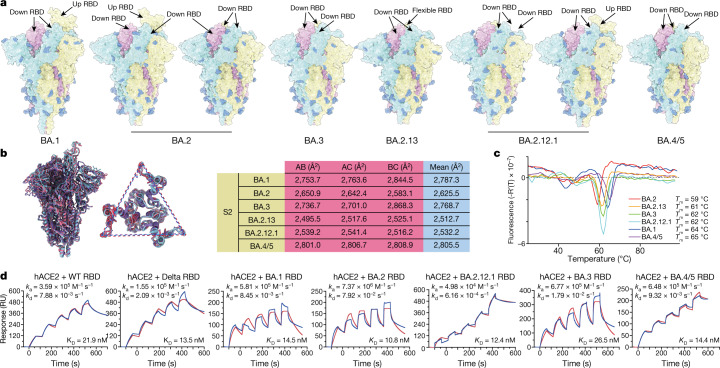
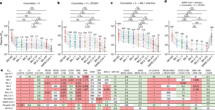
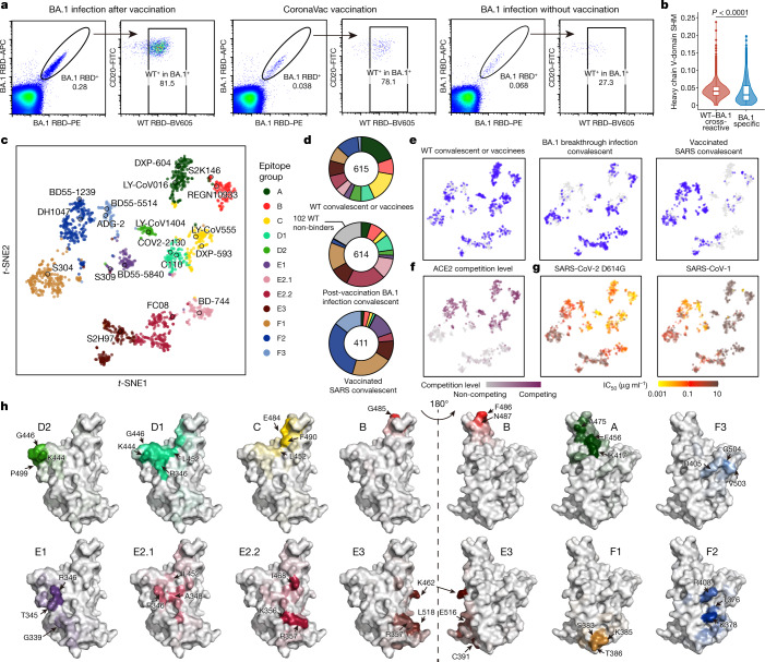
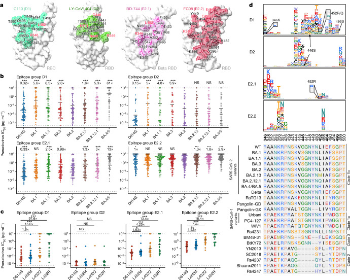
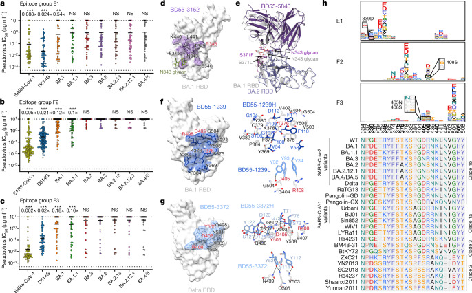
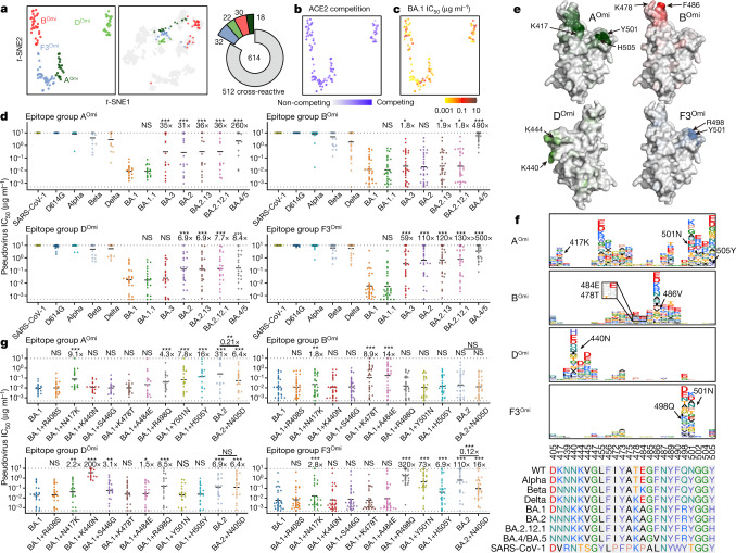
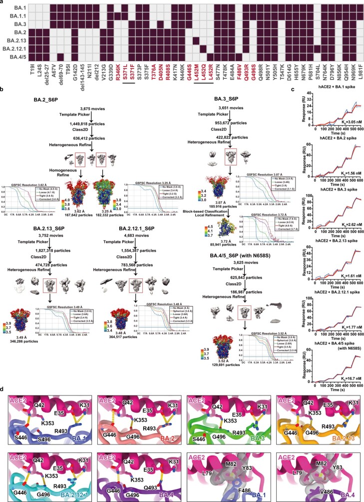
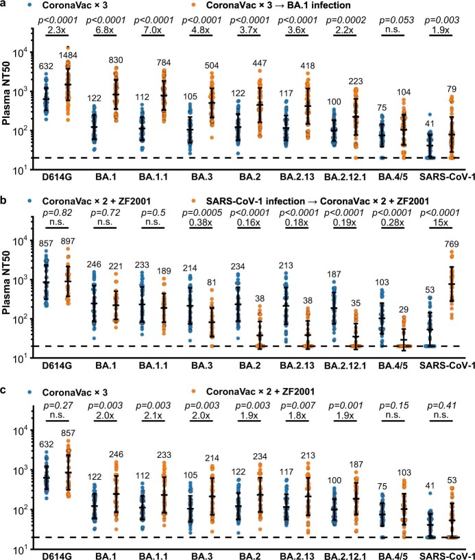
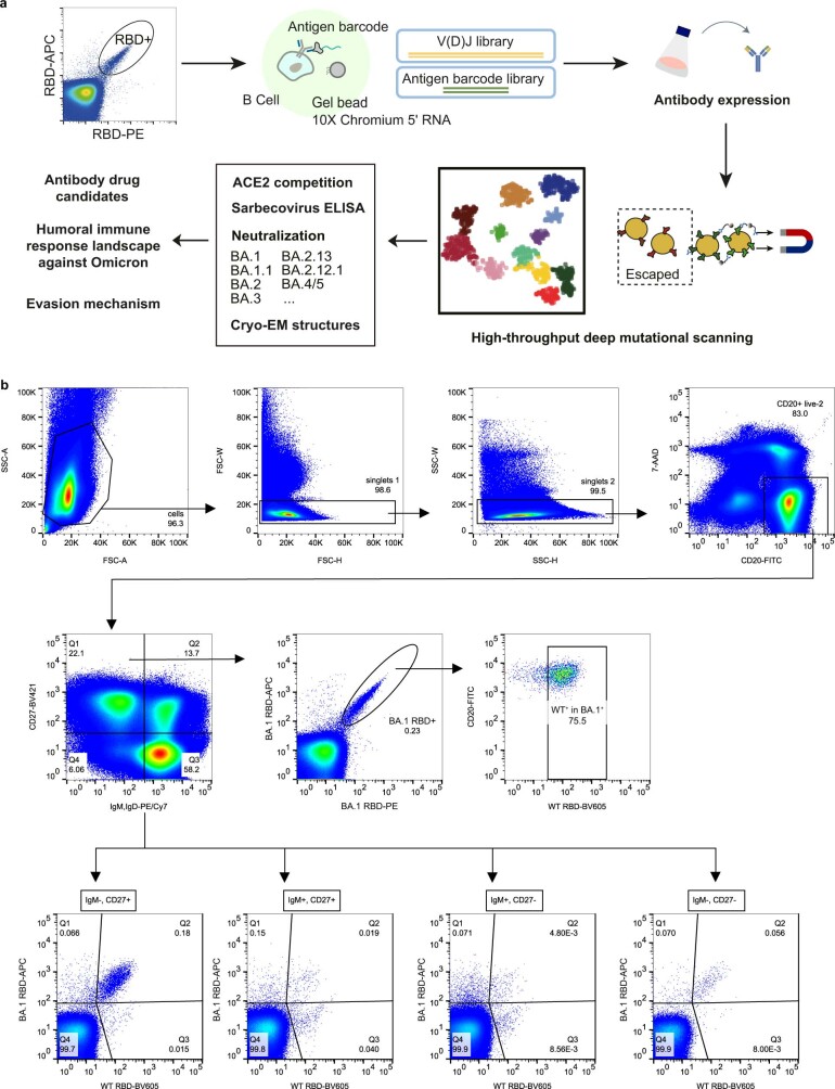
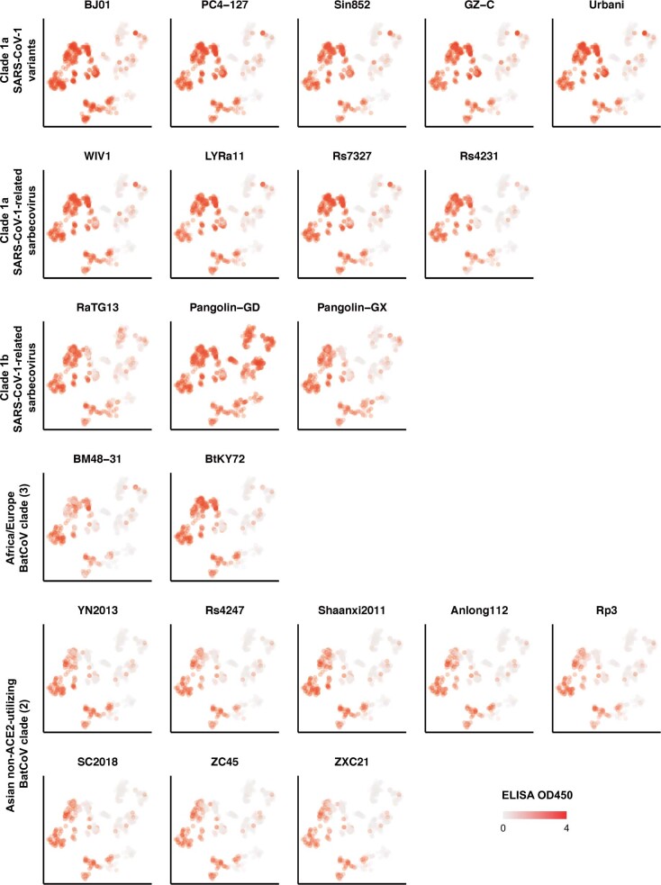
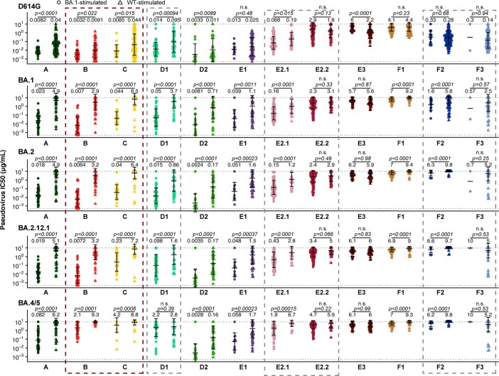
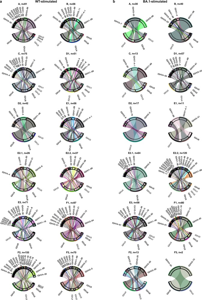
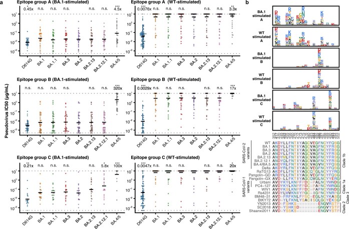
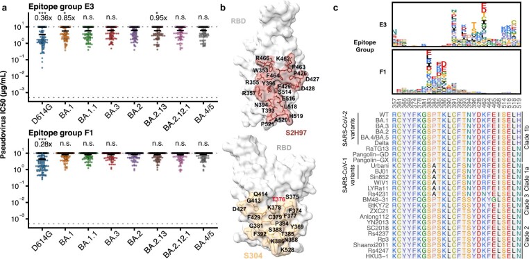
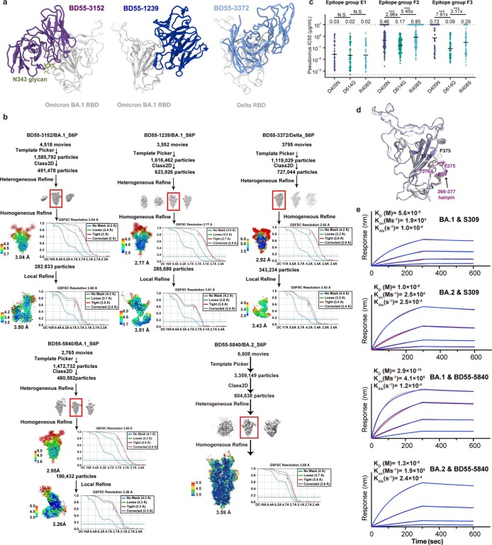
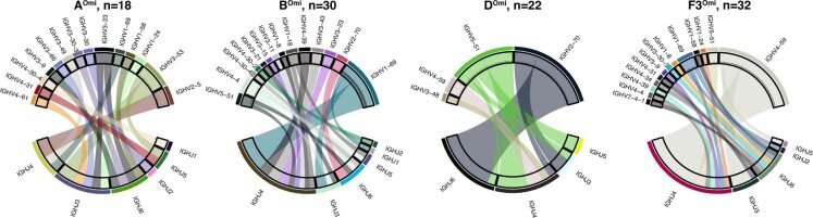
Severe acute respiratory syndrome coronavirus 2 (SARS-CoV-2) Omicron sublineages BA.2.12.1, BA.4 and BA.5 exhibit higher transmissibility than the BA.2 lineage1. The receptor binding and immune-evasion capability of these recently emerged variants require immediate investigation. Here, coupled with structural comparisons of the spike proteins, we show that BA.2.12.1, BA.4 and BA.5 (BA.4 and BA.5 are hereafter referred collectively to as BA.4/BA.5) exhibit similar binding affinities to BA.2 for the angiotensin-converting enzyme 2 (ACE2) receptor. Of note, BA.2.12.1 and BA.4/BA.5 display increased evasion of neutralizing antibodies compared with BA.2 against plasma from triple-vaccinated individuals or from individuals who developed a BA.1 infection after vaccination. To delineate the underlying antibody-evasion mechanism, we determined the escape mutation profiles2, epitope distribution3 and Omicron-neutralization efficiency of 1,640 neutralizing antibodies directed against the receptor-binding domain of the viral spike protein, including 614 antibodies isolated from people who had recovered from BA.1 infection. BA.1 infection after vaccination predominantly recalls humoral immune memory directed against ancestral (hereafter referred to as wild-type (WT)) SARS-CoV-2 spike protein. The resulting elicited antibodies could neutralize both WT SARS-CoV-2 and BA.1 and are enriched on epitopes on spike that do not bind ACE2. However, most of these cross-reactive neutralizing antibodies are evaded by spike mutants L452Q, L452R and F486V. BA.1 infection can also induce new clones of BA.1-specific antibodies that potently neutralize BA.1. Nevertheless, these neutralizing antibodies are largely evaded by BA.2 and BA.4/BA.5 owing to D405N and F486V mutations, and react weakly to pre-Omicron variants, exhibiting narrow neutralization breadths. The therapeutic neutralizing antibodies bebtelovimab4 and cilgavimab5 can effectively neutralize BA.2.12.1 and BA.4/BA.5, whereas the S371F, D405N and R408S mutations undermine most broadly sarbecovirus-neutralizing antibodies. Together, our results indicate that Omicron may evolve mutations to evade the humoral immunity elicited by BA.1 infection, suggesting that BA.1-derived vaccine boosters may not achieve broad-spectrum protection against new Omicron variants.


Conflict of interest statement

X.S.X. and Y. Cao are inventors on the provisional patent applications for BD series antibodies, which includes BD30-604 (DXP-604), BD55-5840 (SA58) and BD55-5514 (SA55). X.S.X. and Y. Cao are founders of Singlomics Biopharmaceuticals. The other authors declare no competing interests.

Figures

Fig. 1
Fig. 1. Structural and receptor-binding characteristics of Omicron subvariants.
a, Surface representation of S-trimers of BA.1, BA.2, BA.3, BA.2.13, BA.2.12.1 and BA.4/BA.5 (BA.4/5) variants. b, Structural interpretation and functional verification of the stability of the spike protein of BA.1, BA.2, BA.3, BA.2.13, BA.2.12.1 and BA.4/BA.5 variants. Left, superimposed structures of spike protein and the S2 domains of BA.1 (purple), BA.2 (red) and BA.4/BA.5 (blue). The binding surface areas between S2 subunits of the variants are calculated in the table on the right. c, Thermoflour analysis for these Omicron variants. Analyses were performed as biological duplicates. d, Binding affinities of RBDs of Omicron variants for hACE2 measured by SPR. Analyses were performed as biological duplicates.
Fig. 2
Fig. 2. BA.2.12.1, BA.4 and BA.5 exhibit stronger antibody evasion than BA.2.
ad, Neutralizing titres against SARS-CoV-2 D614G, Omicron subvariants and SARS-CoV-1 pseudoviruses in plasma from vaccinated and convalescent individuals. a, Individuals who had received 3 doses of CoronaVac (n = 40). b, Individuals who had received 2 doses of CoronaVac and 1 dose of ZF2001 (n = 38). c, Individuals who, after receiving 3 doses of CoronaVac, had been infected with BA.1 and recovered (n = 50). d, People who had recovered from SARS and received 2 doses of CoronaVac and 1 dose of ZF2001 (n = 28). P-values were calculated using two-tailed Wilcoxon signed-rank tests of paired samples. The geometric mean titre is shown above each group of points. e, Neutralizing activity against SARS-CoV-2 variants and sarbecoviruses by therapeutic NAbs. Green, half-maximal inhibitory concentration (IC50) ≤ 30 ng ml−1; white, 30 ng ml−1 < IC50 < 1,000 ng ml−1; red, IC50 ≥ 1,000 ng ml−1; *, IC50 ≥ 10,000 ng ml−1. All neutralization assays were performed as biological duplicates. *P< 0.05, **P< 0.01, ***P< 0.001; NS, not significant (P > 0.05).
Fig. 3
Fig. 3. Isolation, characterization, and comprehensive epitope mapping of SARS-CoV-2 RBD antibodies.
a, FACS analysis of pooled memory B cells (IgMCD27+) from plasma of individuals who have recovered from BA.1 breakthrough infection after vaccination, vaccinated individuals and unvaccinated individuals who have recovered from BA.1 breakthrough infection. The percentage of cells recognizing WT or BA.1 RBD are shown. b, The heavy chain V domain somatic hypermutation (SHM) rate of BA.1-specific (n = 968) and BA.1–WT cross-reactive (n = 4,782) BCRs obtained from 10X scVDJ-seq from individuals who have recovered from BA.1 breakthrough infection after vaccination. Two-tailed Wilcoxon rank-sum test. Boxes show 25th percentile, median and 75th percentile, and violin plots show kernel density estimation curves of the distribution. c, t-SNE and unsupervised clustering of antibodies that bind WT SARS-CoV-2 RBD. Twelve epitope groups were identified on the basis of DMS of 1,538 antibodies. d,e, Epitope distribution and projection of antibodies from plasma of individuals who had recovered from infection with the WT virus, individuals who have recovered from BA.1 breakthrough infection after vaccination, and vaccinated individuals who had recovered from SARS. f, ACE2 competition level determined by competition ELISA (n = 1,286) were projected onto the t-SNE. g, Neutralizing activity against SARS-CoV-2 D614G (n = 1,509) and SARS-CoV-1 (HKU-39849; n = 1,457). h, Average mutational escape score projection of each epitope group on SARS-CoV-2 RBD (Protein Data Bank (PDB): 6M0J). All neutralization assays were performed as biological duplicates.
Fig. 4
Fig. 4. Spike L452 mutants can evade cross-reactive NAbs elicited by BA.1 infection.
a, Epitope of representative antibodies in group D1 (C110; PDB: 7K8V), D2 (LY-CoV1404; PDB: 7MMO), E2.1 (BD-744; PDB: 7EY0) and E2.2 (FC08; PDB: 7DX4). Residues highlighted in red indicate sites that are mutated in Omicron variants. b, Neutralizing activity of NAbs in group D1 (n = 95), D2 (n = 53), E2.1 (n = 90) and E2.2 (n = 161) against spike-pseudotyped SARS-CoV-2 variants. The geometric mean of the fold change in IC50 relative to BA.2 is shown above each plot. Two-tailed Wilcoxon signed-rank test of paired samples, in comparison to IC50 values versus BA.2. c, Neutralizing activity of representative potent NAbs in group D1 (n = 24), D2 (n = 12), E2.1 (n = 23) and E2.2 (n = 23) against SARS-CoV-2 spike L452 mutants. Geometric mean of the fold change in IC50 relative to D614G is shown above each plot. Two-tailed Wilcoxon signed-rank test of paired samples. d, Average escape maps at escape hotspots of antibodies in epitope groups D1, D2, E2.1 and E2.2, and the corresponding multiple sequence alignment of various sarbecovirus RBDs. The height of each amino acid in the escape map represents its mutation escape score. Sites that are mutated in Omicron subvariants are marked in bold. All neutralization assays were performed as biological duplicates.
Fig. 5
Fig. 5. BA.2 subvariants can escape most broad-specificity sarbecovirus-neutralizing antibodies.
ac, Neutralizing activity against SARS-CoV-1 and SARS-CoV-2 subvariants by NAbs in group E1 (a; n = 70), F2 (b; n = 171) and F3 (c; n = 69). The geometric mean of the fold change in IC50 relative to BA.2 is shown above each plot. P-values were calculated using a two-tailed Wilcoxon signed-rank test of paired samples, compared with the IC50 for BA.2. d, The epitope of Group E1 antibody BD55-3152 on the BA.1 RBD. e, Overlay of BD55-5840 in the complex with BA.1 or BA.2 RBD. f,g, The epitope and interactions on the binding interface of BD55-1239 (group F2) (f) and BD55-3372 (group F3) (g). Antibody residues are shown in blue, and RBD residues are in black or red. Residues highlighted in red indicate sites that are mutated in Omicron variants. h, Average escape maps of antibodies in epitope groups E1, F2 and F3, and the corresponding multiple sequence alignment of various sarbecovirus RBDs. The height of each amino acid in the escape map represents its mutation escape score. Sites that are mutated in Omicron subvariants are marked in bold. All neutralization assays were performed as biological duplicates.
Fig. 6
Fig. 6. BA.1-specific antibodies elicited by BA.1 infection exhibit narrow specificity.
a, Four epitope groups were identified among 102 BA.1-specific NAbs via k-means clustering and t-SNE of BA.1 RBD-based DMS profiles. b,c, Distribution of ACE2 competition level (b) and neutralizing activities (c) against BA.1. d, Neutralizing activities of BA.1-specific antibodies against pseudovirus with SARS-CoV-1 and SARS-CoV-2 spike variants (AOmi, n = 18; BOmi, n = 30; DOmi, n = 22; F3Omi, n = 32). The geometric mean of the fold change in IC50 relative to BA.1 is shown above each plot. e, Average mutational escape score projection of each BA.1-specific epitope group on SARS-CoV-2 RBD (PDB: 7WPB). f, Averaged escape maps at escape hotspots of the 102 NAbs in the four epitope groups, and corresponding multiple sequence alignment of various sarbecovirus RBDs. The height of each amino acid in the escape map represents its mutation escape score. Sites that are mutated in Omicron variants are marked in bold. WT-related escaping mutations are highlighted. g, Neutralizing activities of BA.1-specific NAbs against BA.1- or BA.2-based pseudoviruses carrying single substitutions (AOmi, n = 18; BOmi, n = 30; DOmi, n = 22; F3Omi, n = 32). The geometric mean of the fold change in IC50 relative to BA.1 is shown above each plot. Wilcoxon signed-rank test of paired samples, compared with IC50 for BA.1. All neutralization assays were performed as biological duplicates.
Extended Data Fig. 1
Extended Data Fig. 1. Structures and ACE2 binding of emerging Omicron subvariants spike glycoprotein.
a, Mutations on the spike glycoprotein of SARS-CoV-2 Omicron subvariants. Residues that are not identical among Omicron subvariants are colored red. b, Workflow to generate cryo-EM structure of BA.2, BA.3, BA.2.13, BA.2.12.1, BA.4/5 spike glycoprotein trimer with S6P and R683A, R685A substitutions. c, Binding affinities of Omicron variants spike trimers to hACE2 measured by SPR. SPR analyses were conducted in biological duplicates. d, MD simulated interactions between hACE2 and RBD of Omicron variants. Structures of the RBD from Omicron variants and hACE2 are shown as ribbons.
Extended Data Fig. 2
Extended Data Fig. 2. Different immunity backgrounds lead to distinct humoral immunity against Omicron subvariants.
NT50 against SARS-CoV-2, SARS-CoV-1 D614G and Omicron subvariants spike-pseudotyped VSV by plasma samples from a, individuals who received 3 doses CoronaVac with (n = 50) or without (n = 40) BA.1 breakthrough infection; b, individuals who received 2 doses CoronaVac and ZF2001 booster with (n = 28) or without (n = 38) previous SARS-CoV-1 infection; c, individuals who received 3 doses CoronaVac (n = 40) or 2 doses CoronaVac with ZF2001 booster (n = 38). P-values were calculated using two-tailed Wilcoxon rank-sum tests and labeled above the bars. n.s., not significant, p > 0.05. All neutralization assays were conducted in biological duplicates. Geometric means are labeled. Error bars refer to geometric standard deviations.
Extended Data Fig. 3
Extended Data Fig. 3. Workflow for the isolation and characterization of SARS-CoV-2 RBD antibodies.
a, Overall schematic of antibody identification by single cell VDJ sequencing with feature barcodes and epitope analysis by high-throughput deep mutational scanning. b, FACS strategy to enrich BA.1/WT cross-reactive memory B cells or BA.1-specific memory B cells.
Extended Data Fig. 4
Extended Data Fig. 4. ELISA reactivity against 22 sarbecovirus RBD.
Shades of red indicate ELISA OD450 for each antibody against various sarbecoviruses from different clades.
Extended Data Fig. 5
Extended Data Fig. 5. Neutralizing activities of antibodies elicited by SARS-CoV-2 BA.1 or wildtype.
Neutralizing activity against SARS-CoV-2 D614G and Omicron subvariants pseudovirus by antibodies of each epitope group from BA.1 convalescents (BA.1-stimulated. A, n = 30; B, n = 41; C, n = 20; D1, n = 49; D2, n = 17; E1, n = 11; E2.1, n = 64; E2.2, n = 122; E3, n = 57; F1, n = 80; F2, n = 13; F3, n = 2), and from wildtype convalescents or vaccinees (WT-stimulated. A, n = 98; B, n = 55; C, n = 88; D1, n = 46; D2, n = 36; E1, n = 59; E2.1, n = 26; E2.2, n = 39; E3, n = 68; F1, n = 97; F2, n = 158; F3, n = 67). Geometric mean titers (GMT) are annotated above each group of points, and error bars indicate geometric standard deviation. P-values were calculated using two-tailed Wilcoxon rank-sum tests and labeled above the bars. n.s., not significant, p > 0.05. NAbs in the boxed epitope groups showed substantial neutralization potency changes against BA.2.12.1 or BA.4/5 compared to BA.1. All neutralization assays were conducted in biological duplicates.
Extended Data Fig. 6
Extended Data Fig. 6. Heavy chain V-J genes of BA.1-stimulated and WT-stimulated antibodies in each epitope group.
Heavy chain V-J genes combination of a, WT-stimulated antibodies. b, BA.1-stimulated antibodies or each epitope group. The number of NAbs is annotated above the chord plot. IGHV genes are annotated only if the corresponding number of antibodies is greater than one.
Extended Data Fig. 7
Extended Data Fig. 7. Comparison of BA.1-stimulated and WT-stimulated antibodies in group A, B and C.
a, Neutralizing activity against SARS-CoV-2 D614G and Omicron subvariants by BA.1-stimulated (A, n = 30; B, n = 41; C, n = 20) and WT-stimulated (A, n = 98; B, n = 55; C, n = 88) antibodies in Group A, B and C. Geometric mean of IC50 fold changes compared to IC50 against BA.2 are annotated above the bars. P-values were calculated using a two-tailed Wilcoxon signed-rank test of paired samples, in comparison to IC50 against BA.2. *, p < 0.05; **, p < 0.01; ***, p < 0.001; n.s., not significant, p > 0.05. All neutralization assays were conducted in biological duplicates. b, Averaged escape maps at escape hotspots of BA.1-stimulated and WT-stimulated antibodies in group A, B and C, and corresponding MSA of various sarbecovirus RBDs. Height of each amino acid in the escape maps represents its mutation escape score. Mutated sites in Omicron variants are marked in bold.
Extended Data Fig. 8
Extended Data Fig. 8. Antibodies of group E3 and F1 exhibit weak but broad-spectrum neutralization.
a, Neutralizing activity against SARS-CoV-2 D614G and Omicron subvariants by antibodies in group E3 (n = 125) and F1 (n = 177). Geometric mean of IC50 fold changes compared to BA.2 are annotated above the bars. P-values were calculated using a two-tailed Wilcoxon signed-rank test of paired samples, in comparison to IC50 against BA.2. *, p < 0.05; **, p < 0.01; ***, p < 0.001; n.s., not significant, p > 0.05. All neutralization assays were conducted in biological duplicates. b, Epitope of representative antibodies in group E3 (S2H97, PDB: 7M7W) and F1 (S304, PDB: 7JW0). Residues highlighted in red indicate mutated sites in Omicron variants. c, Averaged escape maps at escape hotspots of antibodies in group E3 and F1, and corresponding MSA of various sarbecovirus RBDs. Height of each amino acid in the escape maps represents its mutation escape score. Mutated sites in Omicron variants are marked in bold.
Extended Data Fig. 9
Extended Data Fig. 9. RBD-binding structures and affinity of broad Sarbecovirus antibodies.
a, Cartoon models of Cryo-EM structures of BD55-3152 in complex of BA.1 RBD, BD55-1239 in complex of BA.1 RBD, and BD55-3372 in complex of Delta RBD. b, Workflow to generate refined structural model of BD55-3152 and BD55-1239 in complex of BA.1 RBD, BD55-3372 in complex of Delta RBD, and BD55-5840 in complex of BA.2 RBD. c, Neutralizing activity of representative NAbs in group E1 (n = 68), F2 (n = 139) and F3 (n = 61) against SARS-CoV-2 D614G, in addition to D614G+D405N and D614G+R408S. Geometric mean of IC50 fold changes compared to IC50 against D614G are annotated above the bars. P-values were calculated using a two-tailed Wilcoxon signed-rank test of paired samples. *, p < 0.05; **, p < 0.01; ***, p < 0.001; n.s., not significant, p > 0.05. All neutralization assays were conducted in biological duplicates. d, Conformational comparison between BA.1 and BA.2 RBD regarding the 366-377 hairpin. e, Biolayer interferometry analysis of Group E1 antibodies S309 and BD55-5840 binding to Omicron BA.1 and BA.2 Spike trimer. Biolayer interferometry analyses were conducted in biological duplicates.
Extended Data Fig. 10
Extended Data Fig. 10. HV-HJ gene combination of BA.1-specific antibodies.
Heavy chain V-J gene combination of BA.1-specific neutralizing antibodies in BA.1-specific epitope groups AOmi, BOmi, DOmi and F3Omi.

Comment in

References

    1. Chen C, et al. CoV-spectrum: analysis of globally shared SARS-CoV-2 data to identify and characterize new variants. Bioinformatics. 2021;38:1735–1737. doi: 10.1093/bioinformatics/btab856. - DOI - PMC - PubMed
    1. Greaney AJ, et al. Complete mapping of mutations to the SARS-CoV-2 spike receptor-binding domain that escape antibody recognition. Cell Host Microbe. 2021;29:44–57.e49. doi: 10.1016/j.chom.2020.11.007. - DOI - PMC - PubMed
    1. Cao Y, et al. Omicron escapes the majority of existing SARS-CoV-2 neutralizing antibodies. Nature. 2022;602:657–663. doi: 10.1038/s41586-021-04385-3. - DOI - PMC - PubMed
    1. Westendorf K, et al. LY-CoV1404 (bebtelovimab) potently neutralizes SARS-CoV-2 variants. Cell Rep. 2022;39:110812. doi: 10.1016/j.celrep.2022.110812. - DOI - PMC - PubMed
    1. Zost SJ, et al. Potently neutralizing and protective human antibodies against SARS-CoV-2. Nature. 2020;584:443–449. doi: 10.1038/s41586-020-2548-6. - DOI - PMC - PubMed

Publication types

MeSH terms

Supplementary concepts