. 2022 Jul 5;61(13):1313-1322.
doi: 10.1021/acs.biochem.2c00244. Epub 2022 Jun 17.

Reaction Mechanism and Three-Dimensional Structure of GDP-d-glycero-α-d-manno-heptose 4,6-Dehydratase from Campylobacter jejuni

Affiliations

Reaction Mechanism and Three-Dimensional Structure of GDP-d-glycero-α-d-manno-heptose 4,6-Dehydratase from Campylobacter jejuni

Dao Feng Xiang et al. Biochemistry. .

Abstract

Campylobacter jejuni is a human pathogen and a leading cause of food poisoning in the United States and Europe. Surrounding the outside of the bacterium is a carbohydrate coat known as the capsular polysaccharide. Various strains of C. jejuni have different sequences of unusual sugars and an assortment of decorations. Many of the serotypes have heptoses with differing stereochemical arrangements at C2 through C6. One of the many common modifications is a 6-deoxy-heptose that is formed by dehydration of GDP-d-glycero-α-d-manno-heptose to GDP-6-deoxy-4-keto-d-lyxo-heptose via the action of the enzyme GDP-d-glycero-α-d-manno-heptose 4,6-dehydratase. Herein, we report the biochemical and structural characterization of this enzyme from C. jejuni 81-176 (serotype HS:23/36). The enzyme was purified to homogeneity, and its three-dimensional structure was determined to a resolution of 2.1 Å. Kinetic analyses suggest that the reaction mechanism proceeds through the formation of a 4-keto intermediate followed by the loss of water from C5/C6. Based on the three-dimensional structure, it is proposed that oxidation of C4 is assisted by proton transfer from the hydroxyl group to the phenolate of Tyr-159 and hydride transfer to the tightly bound NAD+ in the active site. Elimination of water at C5/C6 is most likely assisted by abstraction of the proton at C5 by Glu-136 and subsequent proton transfer to the hydroxyl at C6 via Ser-134 and Tyr-159. A bioinformatic analysis identified 19 additional 4,6-dehydratases from serotyped strains of C. jejuni that are 89-98% identical in the amino acid sequence, indicating that each of these strains should contain a 6-deoxy-heptose within their capsular polysaccharides.


Conflict of interest statement

The authors declare no competing conflicts of interests.

Figures
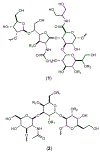
Figure 1.
Figure 1.
Structures of the repeating sugar units in the CPS from C. jejuni strains NCTC 11168 (1) and 81-176 (2). R1 corresponds to the methyl phosphoramidate modification, while R2 represents a methyl ether modification.
Figure 2.
Figure 2.
Sequence similarity network of the 1000 closest homologues to the GMH dehydratase from C. jejuni 81-176. The alignment value cutoff was set to a 68% sequence identity. The sequences from C. jejuni and other Campylobacter species are colored orange and green, respectively. The sequence for the GMH dehydratase from C. jejuni 81-176 is shown as the yellow circle, while that from Y. pseudotuberculosis is shown in blue.
Figure 3:
Figure 3:
UV spectrum of the NAD+ isolated from the purified GMH dehydratase after heat denaturation. Additional details are available in the text.
Figure 4.
Figure 4.
1H NMR spectra of GDP-d-glycero-α-d-manno-heptose (3) and the reaction product after the addition of GMH dehydratase. (A) GDP-d-glycero-α-d-manno-heptose (3). (B) The reaction product, GDP-6-deoxy-4-keto-α-d-lyxo-heptose (4) when the reaction was conducted in D2O. In this product the hydrogen at C5 is deuterated. (C) The reaction product, GDP-6-deoxy-4-keto-α-d-lyxo-heptose (4) when the reaction was conducted in H2O. The hydrogens from the ribose moiety are labelled with an “R”, while those from the manno-heptose moiety are labelled with an “M”. Those resonances labelled with an “*” are from unidentified impurities.
Figure 5.
Figure 5.
Mass spectrometry data for the reactions catalyzed by GMH dehydratase. (A) GDP-d-glycero-α-d-manno-heptose (3) in H2O before the addition of enzyme. (B) GDP-d-glycero-α-d-manno-heptose (3) in D2O before the addition of enzyme. (C) Product of the reaction catalyzed by GMH dehydratase when the reaction was conducted in H2O. (D) Product of the reaction catalyzed by GMH dehydratase when the reaction was conducted in D2O. (E) Product of the reaction catalyzed by Cj1427 (17) when 3 was used as the substrate. The unreacted 3 appears at m/z of 634.07 and the product 7 appears at an m/z of 632.07. (F) Product of the reaction after incubation of GMH dehydratase with the reaction mixture formed from the addition of Cj1427 to 3 in the presence of α-ketoglutarate. Reaction products 8 (from 7) and 4 (from 3) appear at an m/z of 614.07, and 616.07, respectively.
Figure 6:
Figure 6:
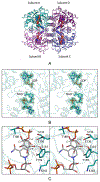
Structure of GMH dehydratase. Shown in (A) is a ribbon drawing of the tetramer as observed in the asymmetric unit. The bound ligands, GDP and NAD(H) are displayed in sphere representations. The observed electron density corresponding to the two ligands is shown in stereo in (B). The electron density map was calculated with (Fo-Fc) coefficients and contoured at 3σ. The ligands were not included in the X-ray coordinate file used to calculate the omit map, and thus there is no model bias. A closeup view of the active site, in stereo, is provided in (C) based on the simple superposition of the substrate onto the structure of GDP-d-mannose 4,6-dehydratase (PDB accession code: 1N7G). The side chains are colored in teal, and the dashed lines indicate possible interactions within 3.2 Å in the modeled GDP-sugar substrate.
Scheme 1:
Scheme 1:
General biosynthetic pathway for the formation of 6-deoxy-heptoses from GDP-d-glycero-α-d-manno-heptose (3) in C. jejuni.
Scheme 2:
Scheme 2:
Coupled enzyme reaction for measuring the kinetic constants for the reaction catalyzed by GMH dehydratase.
Scheme 3:
Scheme 3:
Proposed reaction mechanism for the reaction catalyzed by GMH dehydratase.
Scheme 4:
Scheme 4:
Formation of GDP-d-glycero-4-keto-α-d-lyxo-heptose (7) from GDP-d-glycero-d-manno-heptose (3) catalyzed by Cj1427 in the presence of α-ketoglutarate (α-KG) and the subsequent dehydration of 7 to intermediate 8 catalyzed by GMH dehydratase.
Scheme 5:
Scheme 5:
Proposed reaction mechanism for GDP-d-glycero-d-manno-heptose 4,6-dehydratase.

References

    1. Heimesaat MM, Backert S, Alter T, Bereswill S (2021) Human Campylobacteriosis-a serious infectious threat in a one health perspective. Curr Top Microbiol Immunol. 431, 1–23. - PubMed
    1. Dasti JI, Tareen AM, Lugert R, Zautner AZ, Gross U (2010) Campylobacter jejuni: a brief overview on pathogenicity-associated factors and disease-mediating mechanisms. Int J Med Microbiol. 300, 205–11. - PubMed
    1. Hermans D, Pasmans F, Messens W, Martel A, Immerseel FV, Rasschaert G, Heyndrickx M, Deun KV, Haesebrouck F (2012) Poultry as a host for the zoonotic pathogen Campylobacter jejuni. Vector Borne Zoonotic Dis. 12, 89–98. - PubMed
    1. Humphrey S, Chaloner G, Kemmett K, Davidson N, Williams N, Kipar A, Humphrey T, and Wigley P (2014) Campylobacter jejuni is not merely a commensal in commercial broiler chickens and affects bird welfare. mBio 5, e01364–14. - PMC - PubMed
    1. Facciola A, Riso R, Avventuroso E, Visalli G, Delia SA, and Lagana P (2017) Campylobacter: from microbiology to prevention. J. Prev. Med. Hyg. 58, E79−E92 - PMC - PubMed

Publication types

LinkOut - more resources