Review
. 2022 Sep 14;122(17):13952-13988.
doi: 10.1021/acs.chemrev.1c01035. Epub 2022 Jun 17.

Cryo-electron Microscopic Analysis of Single-Pass Transmembrane Receptors

Affiliations
Review

Cryo-electron Microscopic Analysis of Single-Pass Transmembrane Receptors

Kai Cai et al. Chem Rev. .

Abstract

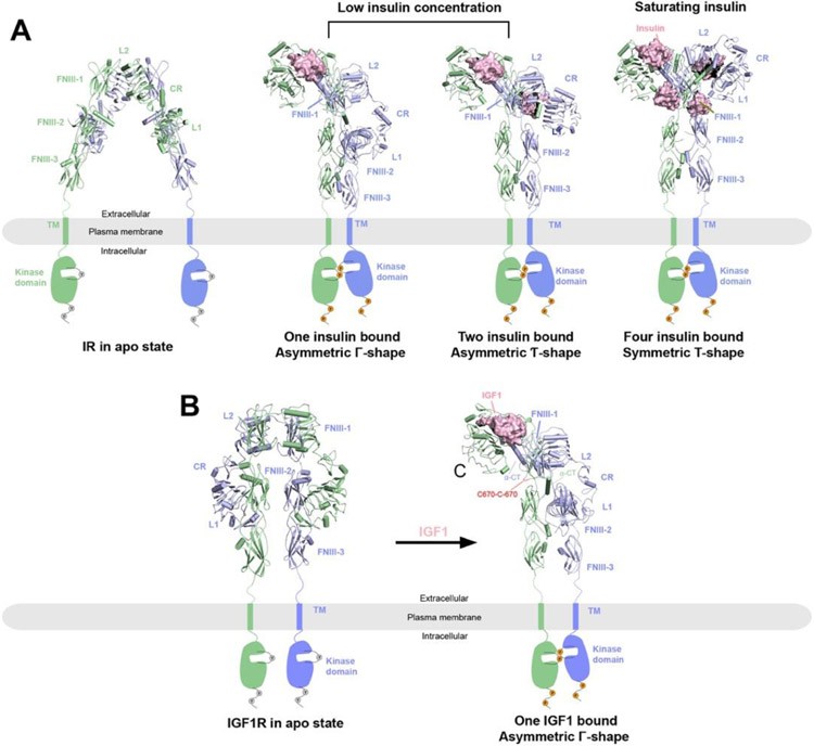
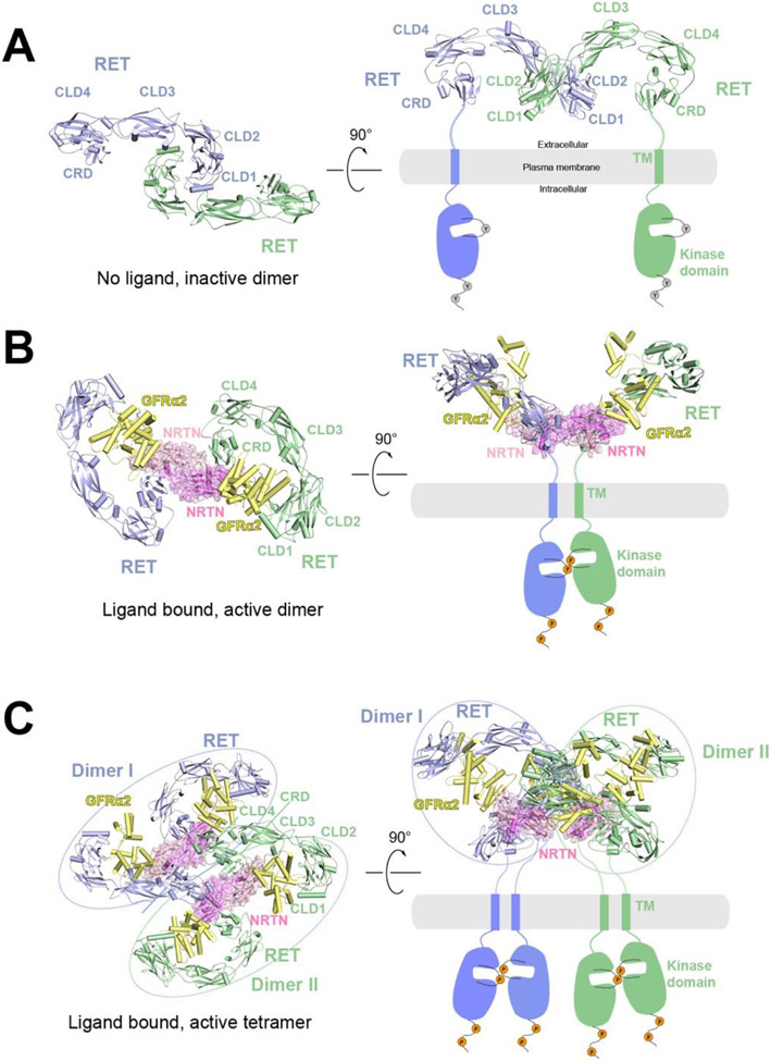
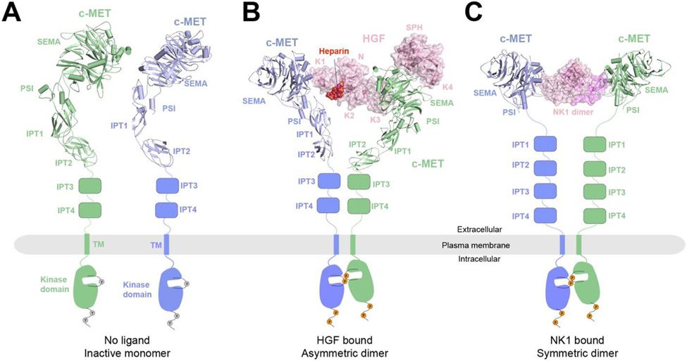
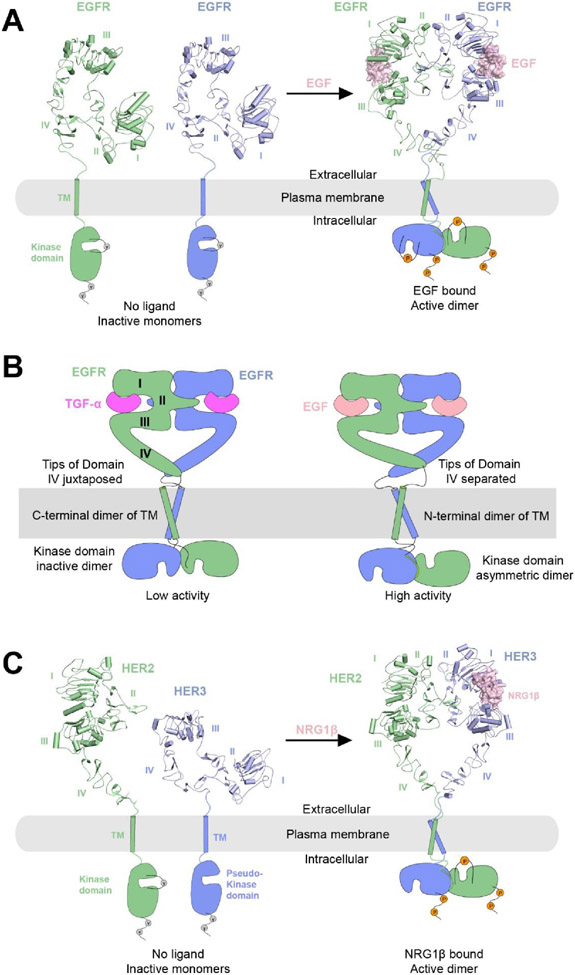
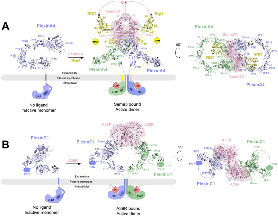
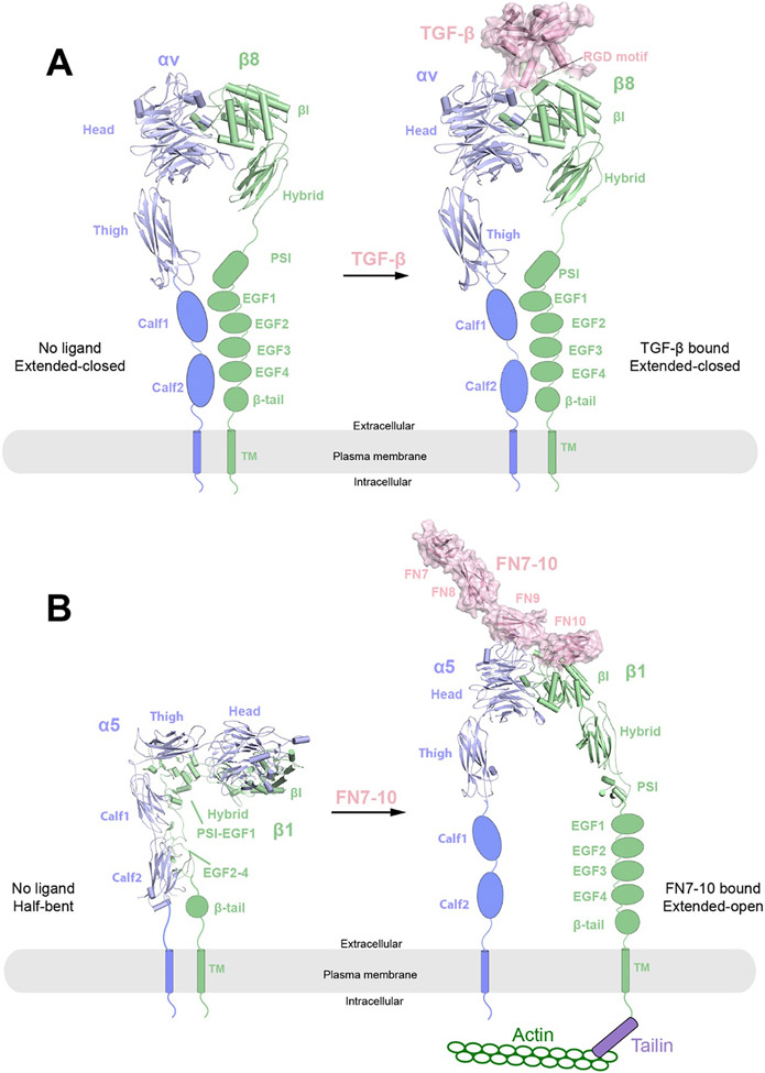
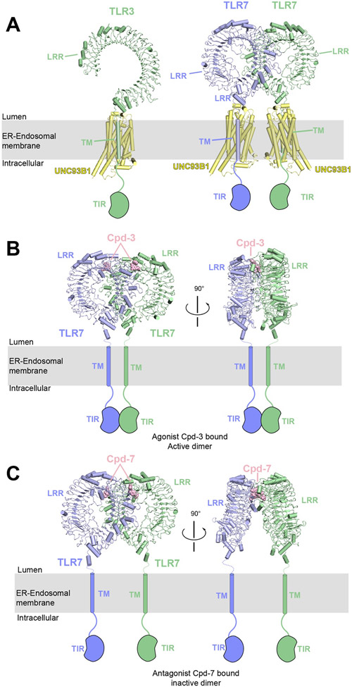
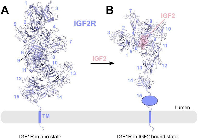
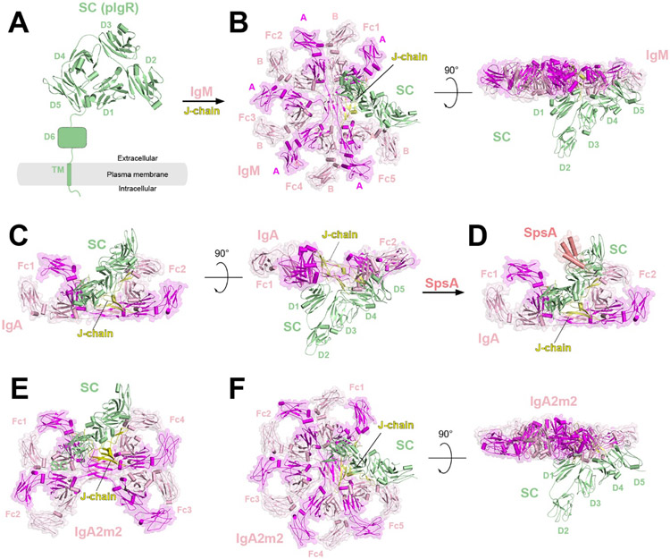
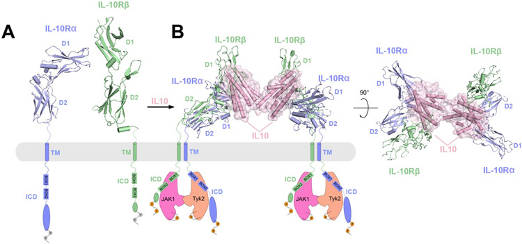
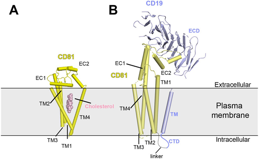
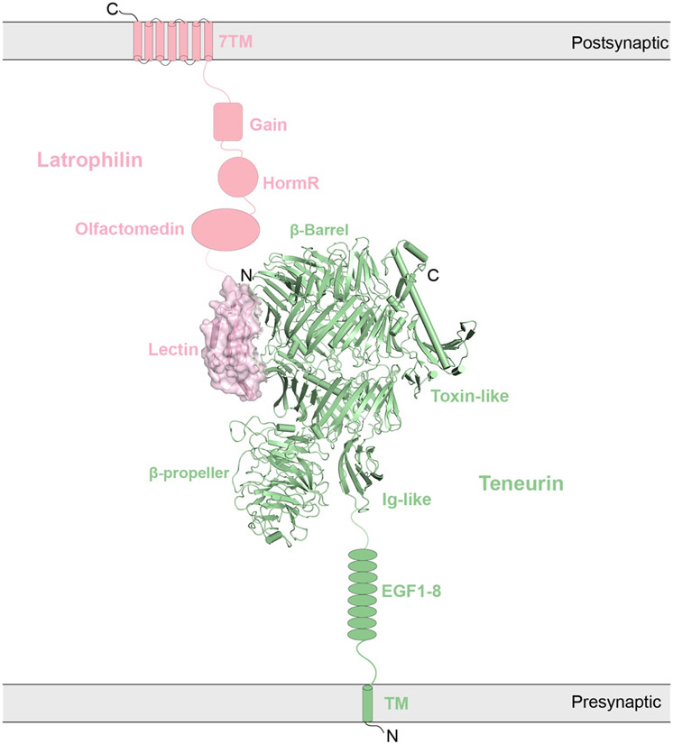
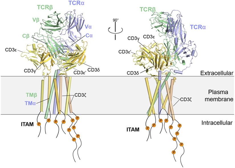
Single-pass transmembrane receptors (SPTMRs) represent a diverse group of integral membrane proteins that are involved in many essential cellular processes, including signal transduction, cell adhesion, and transmembrane transport of materials. Dysregulation of the SPTMRs is linked with many human diseases. Despite extensive efforts in past decades, the mechanisms of action of the SPTMRs remain incompletely understood. One major hurdle is the lack of structures of the full-length SPTMRs in different functional states. Such structural information is difficult to obtain by traditional structural biology methods such as X-ray crystallography and nuclear magnetic resonance (NMR). The recent rapid development of single-particle cryo-electron microscopy (cryo-EM) has led to an exponential surge in the number of high-resolution structures of integral membrane proteins, including SPTMRs. Cryo-EM structures of SPTMRs solved in the past few years have tremendously improved our understanding of how SPTMRs function. In this review, we will highlight these progresses in the structural studies of SPTMRs by single-particle cryo-EM, analyze important structural details of each protein involved, and discuss their implications on the underlying mechanisms. Finally, we also briefly discuss remaining challenges and exciting opportunities in the field.


Figures

Figure 1.
Figure 1.
Overall structures of IR and IGF1R in apo and ligand bounds states reveal activation mechanisms. The two protomers in the receptor dimers are shown in cartoon (blue and green) representations, whereas the ligands are shown in surface (pink) representations. The missing TM and kinase domains in the structures are indicated by schematic drawings. Binding of ligands (insulin or IGF1) leads to conformational changes to the ECD of the receptors, with subsequent dimerization of the TM and kinase domains and activation of the receptors as indicated by the phosphorylation of the kinase domains and C-terminal tails. A) Structures of IR at different states. Left, IR in apo state as an auto-inhibited, Λ-shaped homodimer with lower domains separated (PDB: 4ZXB); middle, full-length 2:1 or 2:2 IR/insulin complexes as Γ- or Ƭ-shaped asymmetric dimers respectively at low insulin concentrations (PDBs: 7STI and 7STJ); right, full-length 2:2 IR/insulin complex as a T-shaped symmetric dimer at saturating insulin concentrations (PDB: 6PXV). B) Left, crystal structure of apo-IGF1R in apo state as an auto-inhibited, Λ-shaped dimer (PDB: 5U8R). Right, cryo-EM structure of full-length 2:1 IGF1R/IGF1 complex (PDB: 6PYH) showing a Γ-shaped asymmetric dimer with two α-CT motifs covalently linked by disulfide bonds.
Figure 2.
Figure 2.
Overall structures of RET at different oligomerization states and proposed activation mechanisms. The protomers in the receptor dimer or tetramer (blue and green) as well as the co-receptor (yellow) are shown in cartoon representations, whereas the ligands are shown in cartoon and surface (pink) representations with semi-transparency. The TM and kinase domains, not present in the structure, are indicated by schematic drawings. A) Structural model of RET in apo state as an inactive dimer in two different views. The structural model is generated based on the crystal structure of CLD1/CLD2 domains (PDB: 2X2U). In this structural model, the two CRD domains are far apart, suggesting an autoinhibited state. B) Cryo-EM structure of the 2:2:2 NRTN/GFRα2/RET complex in two different views (PDB: 6Q2O). In this structure, the two CRD domains are close to each other, consistent with being an active dimer. C) Cryo-EM structure of the 4:4:4 NRTN/GFRα2/RET complex in two different views (PDB: 6Q2R). Dimer I (blue) and Dimer II (green) are structurally identical to the 2:2:2 NRTN/GFRα2/RET complex.
Figure 3.
Figure 3.
Overall structures of c-MET in the apo and different ligand-bounds states and the proposed activation mechanisms. The protomers in the receptor dimer (blue and green) are shown in cartoon representations, whereas the ligands are shown in cartoon and surface (pink) representations with semi-transparency. The domains not present in the structures are represented by schematic drawings. Binding of either HGF or the NK1 dimer leads to dimerization and activation of c-MET receptor. A) Structural model of c-MET in the apo state as inactive monomers. The structural model is taken from the dimer structure of the c-MET/HGF complex (PDB: 7MO7). B) Cryo-EM structure of the c-MET/HGF complex. c-MET forms an asymmetric dimer when bound to full-length HGF and heparin (PDB: 7MO7). Heparin is shown in sphere representation. C) Cryo-EM structure of 2:2 c-MET/NK1 complex showing a symmetric c-MET dimer bridged by the NK1 dimer (PDB: 7MOB).
Figure 4.
Figure 4.
Overall structures of EGFR and HER2-HER3 at their respective apo and ligand-bound states and the proposed activation mechanisms. The protomers in the receptor dimers (blue and green) are shown in cartoon representations, whereas the ligands are shown in cartoon and surface (pink) representations with semi-transparency. The TM and kinase domains are not resolved in the structures, and therefore indicated by schematic representations. Binding of ligands (EGF or NRG1β) leads to conformational changes to EGFR or HER3 that exposes the dimerization interface, enabling the dimerization and activation of receptors. A) Left, crystal structure of apo-EGFR in the inactive monomeric state, in which EGFR shows an autoinhibited configuration with the dimerization interface buried (PDB: 1NQL). Right, cryo-EM structure of the 2:2 EGFR/EGF complex (PDB: 7SYD). B) Schematic representations of the two dimeric conformations of EGFR induced by different type of ligands. Left, the low activity state induced by TGF-α with juxtaposed domain IV, C-terminal TM dimerization and kinase domains forming an inactive dimer. Right, the high activity state induced by EGF, characterized by separated domain IV tips, N-terminal TM dimerization and kinase domains forming an asymmetric dimer. C) Left, crystal structures of HER2 (PDB: 1N8Z) and Her3 (PDB: 1M6B) in their inactive monomeric forms. HER2 lacks the ligand-binding site, and HER3 has a catalytically impaired pseudo-kinase domain. Right, cryo-EM structure of 1:1:1 HER2:HER3:NRG1β complex (PDB: 7MN5).
Figure 5.
Figure 5.
Overall structures of PlexinA4 and PlexinC1 in their respective apo and ligand-bound forms and the proposed activation mechanisms. The protomers in the receptor dimers (blue and green) as well as the coreceptor Nrp1 (yellow) are shown in cartoon representations, whereas the ligands are shown in cartoon and surface (pink) representations with semi-transparency. The missing domains and linkers in each structure are shown as schematic representations. In the apo state, the GAP domains of PlexinA4 or PlexinC1 are in the autoinhibited state with the Rap-binding site inaccessible. Binding of the ligands (Sema3A or A39R) induces plexin dimerization, opening up the GAP active site to allow Rap binding and GTP hydrolysis. A) Left, crystal structure of ECD of PlexinA4 in its apo state (PDB: 5L5K). Middle and right, cryo-EM structure of 2:2:2 PlexinA4/Nrp1/Sema3A complex in two different views (PDB: 7M0R). The structure of the ECD of PlexinA4 in both structures are essentially the same. The lines represent the various linker domains between different protein components that are important to the formation of the complex. The pink lines represent the linker between C723 and the Ig-like domains of Sema3A as well as that between C723 and the C-terminal R770, which engages the binding pocket in the b1 domain of Nrp1. The yellow lines represent the b2-MAM and MAM-TM linkers in Nrp1. B) Left, structural model of monomeric PlexinC1 in its apo state taken from the structure of the PlexinC1/A39R complex (PDB: 6VXK). Middle and right, the cryo-EM structure of the 2:2 PlexinC1/A39R complex (PDB: 6VXK) in two different views.
Figure 6.
Figure 6.
Overall structures of integrins αvβ8 and α5β1 in their respective apo and ligand-bound states. The protomers in the receptor dimers (blue and green) are shown in cartoon representations, whereas the ligands (TGF-β or FN7-10) are shown in cartoon and surface (pink) representations with semi-transparency. The missing domains in each structure are shown as schematic representations. A) Cryo-EM structures of apo state (left) and TGF-β bound state (right) of integrin αvβ8. In both structures, integrin αvβ8 adopts an extended-closed conformation with the leg domains close to each other. B) Cryo-EM structures of apo state (left) and FN7-10 bound state (right) of integrin α5β1. In the apo state, integrin α5β1 adopts a half-bent conformation with the α5 and β1 subunits parallel and leg domains close to each other. In the FN7-10 bound state, integrin α5β1 adopts an extended-open conformation where the leg domains become wide open. The conformational changes allow for the activation of integrin and binding of downstream signaling components such as Talin and Actin.
Figure 7.
Figure 7.
Overall structures of Toll-like receptors TLR3 and TLR7 in complex with UNC93B1 or small molecule agonist/antagonist. The protomers in the TLR receptor dimers (blue and green) as well as UNC93B1 (yellow) are shown in cartoon representations, whereas the ligands (Cpd3 or Cpd7) are shown as spheres. The missing domains (TM and TIR) in each structure are shown as schematic representations. A) Cryo-EM structure of 1:1 TLR3/UNC93B1 (left, PDB: 7C76) and 2:2 TLR7/UNC93B1 (right, PDB: 7CYN) complexes. B-C) Cryo-EM structure of 2:2 TLR7/Cpd3 (PDB: 6LVZ) and 2:2 TLR7/Cpd7 (PDB: 6LW1). Cpd3 is an agonist for TLR7, which induces the formation of closed dimer, promoting TIR dimerization and TLR7 activation. In contrast, Cpd7 is an antagonist for TLR7, which induces the formation of open dimer, consequently inhibiting TIR dimerization and TLR7 activation.
Figure 8.
Figure 8.
Overall structure of full-length IGF2R in the apo and IGF2-bound states. A) Cryo-EM structure of apo-IGF2R (PDB: 6UM1). The 15 domains in ECD are organized into an elongated helix-like assembly. B) Cryo-EM structure of the 1:1 IGF2R/IGF2 complex (PDB: 6UM2). Domains 1-14 rearrange into a pistol-like conformation. Domain 15 becomes disordered and is missing in the EM density. IGF2R (blue) is shown in cartoon representation, whereas the ligand IGF2 is shown in surface representation with semi-transparency. The missing domains in each structure are shown as schematic representations.
Figure 9.
Figure 9.
Overall structures of pIgR (SC) in apo and ligand bound states. pIgR receptor (SC, green) and J-chain (yellow) are shown in cartoon representations, whereas the ligands (IgM, IgA and IgA2m2) are shown in surface representation with semi-transparency. A) Crystal structure of SC in its apo state (PDB: 5D4K), in which SC adopts a closed triangle-shaped structure with D1 and D5 close to each other. The missing D6 and TM domains of pIgR are shown as schematic representations. B) Cryo-EM structure of the pentameric IgM in complex with J-chain and SC (SIgM), in which the D2-D5 domains of SC rearrange into a liner conformation (PDB:7K0C). The subunits A and B for each homodimer are defined based on the previous literature. C) Cryo-EM structure of dimeric SIgA complex (PDB: 6LX3) in two different views, which shows a boomerang-like structure with D2-D5 domains of SC adopting a liner conformation, similar to that of SIgM. D) Cryo-EM structure of dimeric SIgA in complex with SpsA. The structure of SIgA remains unchanged while SpsA binds to the D3-D4 junction of SC. E-F) Cryo-EM structures of tetrameric (E) and pentameric (F) SIgA2m2 complexes.
Figure 10.
Figure 10.
Overall structure of IL-10Rα and IL-10Rβ complex in apo or IL-10 bound states and the proposed activation mechanism. Receptors IL-10Rα (blue) and IL-10Rβ (green) are shown in cartoon representations, whereas the ligand IL-10 is shown as surface representations with semi-transparency. The missing domains (TM and TIR) in each structure are shown as schematic representations. A) Crystal structures of IL-10Rα and IL-10Rβ in their respective apo states as inactive monomers (PDBs: 1Y6K and 3LQM). The ECDs of both proteins consist of D1 and D2 domains. IL-10Rα has a longer ICD than IL-10Rβ, both contains two regions (named Box1 and Box2) that interact with the JAK1 or Tyk2 kinases. B) Cryo-EM structure of 2:2:2 IL-10/IL-10Rα/IL-10Rβ in two different views (PDB: 6X93). Dimerization of IL-10Rα/IL-10Rβ induced by IL-10 leads to engagement of JAK1 and Tyk2 kinases, and phosphorylation of JAK1 and Tyk2 as well as the ICDs of IL-10Rα and IL-10Rβ.
Figure 11.
Figure 11.
Cartoon representations of the overall structures of tetraspanin CD81 alone and in complex with the B-cell coreceptor CD19. A) Crystal structure of CD81 (PDB: 5TCX) reveals a cholesterol binding pocket. B) Cryo-EM structure of the CD81/CD19 complex (PDB: 7JIC). The CTD of CD19 and the flexible linker between CTD of CD19 and TM1 of CD81 are shown as schematic representations.
Figure 12.
Figure 12.
Cryo-EM structure of 1:1 Teneurin (TEN2)/Latrophilin (LPHN3) complex (PDB: 6VHH). Four domains of TEN2 (Toxin-like, β-propeller, β-Barrel, and Ig-like) and one domain of LPHN3 (Lectin) are shown in the structure. The domains that are missing in the structure are shown as schematic representations. TEN2 (green, shown in cartoon representation) is localized to the presynaptic membrane, whereas LPHN3 (pink, shown in surface representation with semi-transparency) is localized to the postsynaptic membrane.
Figure 13.
Figure 13.
Cryo-EM structure of the TCR-CD3 holo-complex. CD3 contains 4 chains- δ, γ, ε and ζ that forms three dimers. Each of δ, γ and ε chains contains an ECD, a TM and an ICD contains an immunoreceptor-tyrosine-based activation motif (ITAM), whereas CD3ζ contains a short extracellular loop, a TM and an ICD with three ITAMs. The TCRα/TCRβ/CD3 complex is assembled by the TCRαβ, CD3γε, CD3δε, and CD3ζζ dimers in a 1:1:1:1 stoichiometry. Activation of the complex by the antigenic peptide/MHC complex leads to phosphorylation of ITAMs of CD3.

References

    1. Almen MS; Nordstrom KJ; Fredriksson R; Schioth HB Mapping the human membrane proteome: a majority of the human membrane proteins can be classified according to function and evolutionary origin. BMC Biol. 2009, 7, 50. - PMC - PubMed
    1. Reis R; Moraes I Structural biology and structure-function relationships of membrane proteins. Biochem. Soc. Trans 2018, DOI:10.1042/BST20180269 10.1042/BST20180269. - DOI - DOI - PubMed
    1. Engel A; Gaub HE Structure and mechanics of membrane proteins. Annu. Rev. Biochem 2008, 77, 127–148. - PubMed
    1. Overington JP; Al-Lazikani B; Hopkins AL How many drug targets are there? Nat. Rev. Drug Discov 2006, 5, 993–996. - PubMed
    1. Bugge K; Lindorff-Larsen K; Kragelund BB Understanding single-pass transmembrane receptor signaling from a structural viewpoint-what are we missing? FEBS J. 2016, 283, 4424–4451. - PubMed

Publication types

Substances