. 2022 Jun 17;12(1):10201.
doi: 10.1038/s41598-022-14158-1.

Curcumin ameliorated low dose-Bisphenol A induced gastric toxicity in adult albino rats

Affiliations

Curcumin ameliorated low dose-Bisphenol A induced gastric toxicity in adult albino rats

Omnia Ibrahim Ismail et al. Sci Rep. .

Abstract

Bisphenol A (BPA) is one of the most common worldwide chemicals involved in the industry of polycarbonate plastics, medical devices, and pharmaceuticals. Forty three-month-old albino rats were randomly classified into four groups. Group Ӏ received a daily corn oil dose (5 mL/kg/ body weight, BW) through a gastric tube for one month, Group ӀӀ received a daily dose of Curcumin (200 mg/kg body weight (B.W.) through a gastric tube for one month, Group ӀӀӀ received a daily dose of BPA (0.5 μg/kg B.W.) through a gastric tube for one month and Group ӀV received concomitant daily doses of Bisphenol A and Curcumin as the regimen described in groups ӀӀ and ӀӀӀ. The rats were sacrificed, and glandular portion of stomach was dissected and processed for light, immunohistochemical and ultrastructural study. BPA induced destructed gastric glands, dilated congested blood vessels, submucosal oedema, decreased PAS-positive reactivity, increased collagen fibres deposition, decrease in the positive BCL2 immunoexpression, increased positive PCNA immunoexpression, reduction in the gastric mucosal height and destructive changes in the enteroendocrine, chief and parietal cells. Curcumin coadministration provoked an obvious improvement in the gastric structure. BPA exposure has toxic effects on the glandular portion of the stomach in rats. Otherwise, Curcumin coadministration has exhibited protective impact on the architecture of the stomach.


Conflict of interest statement

The authors declare no competing interests.

Figures

Figure 1
Figure 1
The photomicrographs of the sections in the glandular portion of the stomach in (a) the group Ӏ showing the gastric mucosa composed of the parietal cells (arrow) having a central spherical nucleus and an acidophilic cytoplasm. Small Chief cells (tailed arrow) have basal nuclei and an apical blue-purple stained cytoplasm. (b) the group ӀӀ showing the gastric mucosa composed of the parietal cells (arrow) and Chief cells (tailed arrow) that appear more or less than in the group Ӏ. (c) the group ӀӀӀ showing destructed gastric glands (arrow head) with vacuolations (curved arrow) and dilated congested blood vessels (bv). The parietal cells have dense nuclei and cytoplasm(arrow). Chief cells have a vacuolated cytoplasm (tailed arrow). (d) the group ӀӀӀ showing dilated congested blood vessels (bv). Submucosal oedema is observed (asterisk). (e) the group ӀV showing some parietal cells have a vacuolated cytoplasm (arrow). Other parietal cells (arrowhead) and Chief cells (tailed arrow) appear more or less than normal. Hx & E, × 400, scale bar = 20 µm).
Figure 2
Figure 2
The photomicrographs of the sections in the glandular portion of the stomach in (a) the group Ӏ showing a strong positive Periodic Acid Schiff (PAS) reaction in the lining epithelium and neck cells (arrow) is noticed. (b) the group ӀӀ showing a strong positive PAS reaction (arrow) more or less than in group Ӏ. (c) the group ӀӀӀ showing a decrease in PAS reaction in the lining epithelium and neck cells (arrow). (d) the group ӀV showing a restoration of the positive PAS reaction in the lining epithelium and neck cells (arrow). (PAS, × 200, scale bar = 50 µm).
Figure 3
Figure 3
The photomicrographs of the sections in the glandular portion of the stomach in (a) the group Ӏ showing a normal distribution of the collagen fibres among the bases of the gastric glands, in the lamina propria and interstitial between the glands (arrow). (b) the group ӀӀ showing the collagen fibres distribution (arrow) more or less than the group Ӏ. (c) the group ӀӀӀ showing an increased collagen fibres deposition (arrow). (d) the group ӀV showing a minimal interstitial collagen fibres deposition (arrow). (Masson’s Trichrome, × 200, scale bar = 50 µm).
Figure 4
Figure 4
The photomicrographs of the sections in the glandular portion of the stomach in (a) the group Ӏ showing a positive BCL2 immunoexpression (arrow) in the gastric glands. (b) the group ӀӀ showing a positive BCL2 immunoexpression (arrow) more or less than the group Ӏ. (c) the group ӀӀӀ showing an apparent decrease in the positive BCL2 immunoexpression (arrow) as compared to the group Ӏ (d) the group ӀV showing an apparent increase in the positive BCL2 immunoexpression (arrow) as compared to the group ӀӀӀ. (BCL2 immunohistochemical staining × 200, Scale bar = 50 µm).
Figure 5
Figure 5
The photomicrographs of the sections in the glandular portion of the stomach in (a) the group Ӏ showing a positive PCNA immunoexpression (arrow) in the gastric cells’ nuclei of the upper gastric glands. (b) the group ӀӀ showing a positive PCNA immunoexpression(arrow) more or less than the group Ӏ. (c) the group ӀӀӀ showing an apparently increased positive PCNA immunoexpression (arrow)in the gastric cells’ nuclei extending into the basal part of the gastric glands as compared to the group Ӏ (d) the group ӀV showing an apparent decrease in the positive PCNA immunoexpression (arrow) in the gastric cells’ nuclei as compared to the group ӀӀӀ. (PCNA immunohistochemical staining × 200, Scale bar = 50 µm).
Figure 6
Figure 6
The photomicrographs of the sections in the glandular portion of the stomach in (a, b, c, d) showing a negative EGFR immunoexpression in all studied groups; Ӏ, ӀӀ, ӀӀӀ, ӀV, receptively. (EGFR immunohistochemical staining × 200, Scale bar = 50 µm).
Figure 7
Figure 7
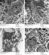
The electron photomicrographs of the enteroendocrine cell in (a) the group Ӏ showing an euchromatic nucleus (N) with many tiny electron-dense granules (G) and abundant ribosomes (RI). (b) the group ӀӀ showing a similar ultrastructural morphology to that in the group Ӏ. (c) the group ӀӀӀ showing an electron dense nucleus (N) and many vacuolations (V). Few tiny electron-dense granules (G) and a loss of cell organelles are noticed. A pyknotic shrunken nucleus (arrow) is seen. (d) the group ӀV showing an euchromatic nucleus (N) with a nuclear membrane indentation (arrow). The cytoplasm shows many tiny electron-dense granules (G) and vacuolations (V). (TEM × 5800, Scale bar = 2 µm).
Figure 8
Figure 8
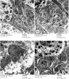
The electron photomicrographs of the Chief cell in (a) the group Ӏ showing an euchromatic nucleus (N), mitochondria (M) and an abundant packed rough endoplasmic reticulum (R). (b) the group ӀӀ showing an ultrastructural morphology as in the group Ӏ. (c) the group ӀӀӀ showing an electron dense nucleus (N), electron dense mitochondria (M) and many vacuolations (V). Dilatation and elongation of the rough endoplasmic reticulum (R) are observed. (d) the group ӀV showing an electron dense nucleus (N) with nucleolus(nu) and a dilated rough endoplasmic reticulum (R). (TEM × 5800, Scale bar = 2 µm).
Figure 9
Figure 9
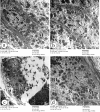
The electron photomicrographs of the parietal cell in (a) the group Ӏ showing an oval euchromatic nucleus (N). The cytoplasm contains abundant mitochondria (M), intracellular canaliculi (IC) and free ribosomes (RI). (b) the group ӀӀ showing an ultrastructural morphology as in the group Ӏ. (c) the group ӀӀӀ showing a marked loss of cell organelles with a vacuolated cytoplasm (V). Few electron dense mitochondria (M) and another destructed one (arrow) are noticed. The euchromatic nucleus (N) is seen. (d) the group ӀV showing an oval euchromatic nucleus (N). The cytoplasm contains the intracellular canaliculi (IC) and free ribosomes (RI). Some mitochondria (M) appear normal; however, others are electron dense (arrow). (TEM × 5800, Scale bar = 2 µm).

Similar articles

Cited by

References

    1. Bahey NG, Abd Elaziz HO, Gadalla KKE. Potential toxic effect of bisphenol A on the cardiac muscle of adult rat and the possible protective effect of Omega-3: a histological and immunohistochemical study. J. Microsc. Ultrastruct. 2019;7(1):1. doi: 10.4103/JMAU.JMAU_53_18. - DOI - PMC - PubMed
    1. Abdelzaher W, Ali D, Khalil W. Could Licorice prevent bisphenol a-induced biochemical, histopathological and genetic effects in the adult male Albino rats? Ain Shams J. Forensic Med. Clin. Toxicol. 2018;30(1):73–87. doi: 10.21608/ajfm.2018.18198. - DOI
    1. Kanwal Q, Qadir A, Iqbal HH, Munir B. Healing potential of Adiantum capillus-veneris L. plant extract on bisphenol A-induced hepatic toxicity in male albino rats. Environ. Sci. Pollut. Res. 2018;25(12):11884–11892. doi: 10.1007/s11356-018-1211-3. - DOI - PubMed
    1. El Henafy HM, Ibrahim MA, Abd El Aziz SA, Gouda EM. Oxidative Stress and DNA methylation in male rat pups provoked by the transplacental and translactational exposure to bisphenol A. Environ. Sci. Pollut. Res. 2020;27(4):4513–4519. doi: 10.1007/s11356-019-06553-5. - DOI - PubMed
    1. Yousaf B, Liu G, Wang R, Qadir A, Ali MU, Kanwal Q, Munir B, Abbas Z. Bisphenol A exposure and healing effects of Adiantum capillus-veneris L. plant extract (APE) in bisphenol A-induced reproductive toxicity in albino rats. Environ. Sci. Pollut. Res. 2016;23(12):11645–11657. doi: 10.1007/s11356-016-6330-0. - DOI - PubMed

Publication types