

FAM111A is dispensable for electrolyte homeostasis in mice
- PMID: 35715480
- PMCID: PMC9205974
- DOI: 10.1038/s41598-022-14054-8
FAM111A is dispensable for electrolyte homeostasis in mice
Abstract
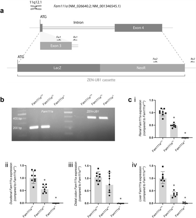
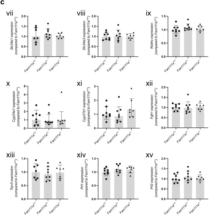
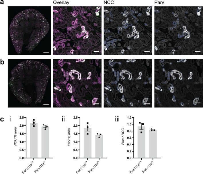
Autosomal dominant mutations in FAM111A are causative for Kenny-Caffey syndrome type 2. Patients with Kenny-Caffey syndrome suffer from severe growth retardation, skeletal dysplasia, hypoparathyroidism, hypocalcaemia, hyperphosphataemia and hypomagnesaemia. While recent studies have reported FAM111A to function in antiviral response and DNA replication, its role in regulating electrolyte homeostasis remains unknown. In this study, we assessed the role of FAM111A in the regulation of serum electrolyte balance using a Fam111a knockout (Fam111a-/-) C57BL/6 N mouse model. Fam111a-/- mice displayed normal weight and serum parathyroid hormone (PTH) concentration and exhibited unaltered magnesium, calcium and phosphate levels in serum and 24-hour urine. Expression of calciotropic (including Cabp28k, Trpv5, Klotho and Cyp24a1), magnesiotropic (including Trpm6, Trpm7, Cnnm2 and Cnnm4) and phosphotropic (Slc20a1, Slc20a2, Slc34a1 and Slc34a3) genes in the kidneys, duodenum and colon were not affected by Fam111a depletion. Only Slc34a2 expression was significantly upregulated in the duodenum, but not in the colon. Analysis of femurs showed unaffected bone morphology and density in Fam111a-/- mice. Kidney and parathyroid histology were also normal in Fam111a-/- mice. In conclusion, our study is the first to characterise the function of FAM111A in vivo and we report that mice lacking FAM111A exhibit normal electrolyte homeostasis on a standard diet.
© 2022. The Author(s).
Conflict of interest statement
The authors declare no competing interests.
Figures


Similar articles
-
Disruption of the c-terminal serine protease domain of Fam111a does not alter calcium homeostasis in mice.Physiol Rep. 2024 May;12(9):e15977. doi: 10.14814/phy2.15977. Physiol Rep. 2024. PMID: 38697929 Free PMC article.
-
A recurrent de novo FAM111A mutation causes Kenny-Caffey syndrome type 2.J Bone Miner Res. 2014 Apr;29(4):992-8. doi: 10.1002/jbmr.2091. J Bone Miner Res. 2014. PMID: 23996431
-
FAM111A mutations result in hypoparathyroidism and impaired skeletal development.Am J Hum Genet. 2013 Jun 6;92(6):990-5. doi: 10.1016/j.ajhg.2013.04.020. Epub 2013 May 16. Am J Hum Genet. 2013. PMID: 23684011 Free PMC article.
-
[Kenny-Caffey syndrome and its related syndromes].Nihon Rinsho. 2015 Nov;73(11):1959-64. Nihon Rinsho. 2015. PMID: 26619675 Review. Japanese.
-
Case report: Late middle-aged features of FAM111A variant, Kenny-Caffey syndrome type 2-suggestive symptoms during a long follow-up.Front Endocrinol (Lausanne). 2023 Jan 4;13:1073173. doi: 10.3389/fendo.2022.1073173. eCollection 2022. Front Endocrinol (Lausanne). 2023. PMID: 36686468 Free PMC article. Review.
Cited by
-
Quantitative hypermorphic FAM111A alleles cause autosomal recessive Kenny-Caffey syndrome type 2 and osteocraniostenosis.JCI Insight. 2025 Feb 11;10(6):e186862. doi: 10.1172/jci.insight.186862. eCollection 2025 Mar 24. JCI Insight. 2025. PMID: 39932783 Free PMC article.
-
Disruption of the c-terminal serine protease domain of Fam111a does not alter calcium homeostasis in mice.Physiol Rep. 2024 May;12(9):e15977. doi: 10.14814/phy2.15977. Physiol Rep. 2024. PMID: 38697929 Free PMC article.
-
Gene-nutrient interactions that impact magnesium homeostasis increase risk for neural tube defects in mice exposed to dolutegravir.Front Cell Dev Biol. 2023 Jun 12;11:1175917. doi: 10.3389/fcell.2023.1175917. eCollection 2023. Front Cell Dev Biol. 2023. PMID: 37377737 Free PMC article.
-
Functions and evolution of FAM111 serine proteases.Front Mol Biosci. 2022 Dec 15;9:1081166. doi: 10.3389/fmolb.2022.1081166. eCollection 2022. Front Mol Biosci. 2022. PMID: 36589246 Free PMC article. Review.
-
Alteration in expression and subcellular localization of the androgen receptor- regulated FAM111A protease is associated with emergence of castration resistant prostate cancer.Neoplasia. 2025 Aug;66:101181. doi: 10.1016/j.neo.2025.101181. Epub 2025 May 29. Neoplasia. 2025. PMID: 40446667 Free PMC article.
References
Publication types
MeSH terms
Substances
LinkOut - more resources
Full Text Sources
Molecular Biology Databases
Miscellaneous
