. 2022 Sep;127(5):824-835.
doi: 10.1038/s41416-022-01865-9. Epub 2022 Jun 17.

Selective cell cycle arrest in glioblastoma cell lines by quantum molecular resonance alone or in combination with temozolomide

Affiliations

Selective cell cycle arrest in glioblastoma cell lines by quantum molecular resonance alone or in combination with temozolomide

Daniela Catanzaro et al. Br J Cancer. 2022 Sep.

Abstract

Background: Glioblastoma is the most aggressive form of brain cancer, characterised by high proliferation rates and cell invasiveness. Despite advances in surgery and radio-chemotherapy, patients continue to have poor prognoses, with a survival rate of 14-15 months. Thus, new therapeutic strategies are needed. Non-ionising electromagnetic fields represent an emerging option given the potential advantages of safety, low toxicity and the possibility to be combined with other therapies.

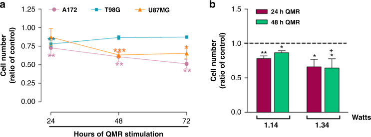
Methods: Here, the anticancer activity of quantum molecular resonance (QMR) was investigated. For this purpose, three glioblastoma cell lines were tested, and the QMR effect was evaluated on cancer cell proliferation rate and aggressiveness. To clarify the QMR mechanism of action, the proteomic asset after stimulation was delineated. Mesenchymal stromal cells and astrocytes were used as healthy controls.

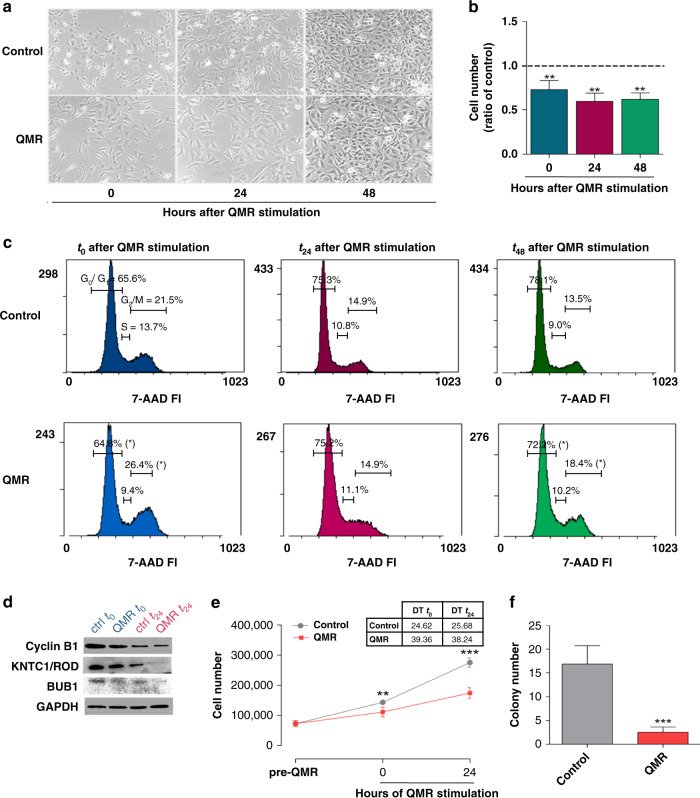
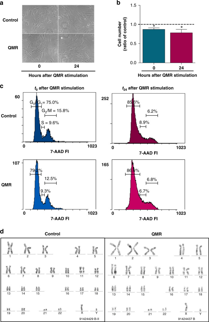
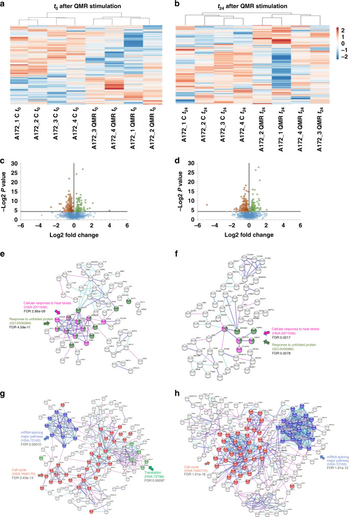
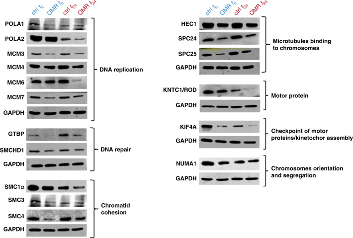
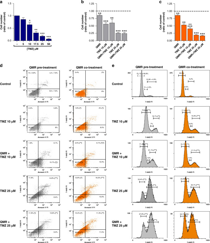
Results: QMR affected cancer cell proliferation, inducing a significant arrest of cell cycle progression and reducing cancer tumorigenicity. These parameters were not altered in healthy control cells. Proteomic analysis suggested that QMR acts not only on DNA replication but also on the machinery involved in the mitotic spindle assembly and chromosome segregation. Moreover, in a combined therapy assessment, QMR significantly enhanced temozolomide efficacy.

Conclusions: QMR technology appears to be a promising tool for glioblastoma treatment.


Conflict of interest statement

DC, GM, AB, MB, KC, MM, AM, PC, RB, DP, FAR, LP, MR and GA declare no competing interests. GP is the cofounder and AP is an employee of Telea Electronic Engineering srl, Sandrigo, Italy. They had no role in the design, execution, interpretation or writing of the study.

Figures

Fig. 1
Fig. 1. Effect of QMR stimulation on A172 glioblastoma cells.
a Representative images of A172 at 0, 24 and 48 h after QMR exposure, acquired with an Axiovert 40 CFL inverted light microscope (Carl Zeiss, Oberkochen, Germany; ×10). b Cell viability was detected by trypan blue exclusion assay. Histograms represent the ratio of viable cells relative to that in unstimulated cells. c Cell cycle progression at 0, 24, and 48 h after QMR stimulation was monitored by flow cytometry. Representative cytograms report the percentage of cells in each cell cycle phase (G0–G1, S, G2–M). d Protein expression of cell cycle key regulators was evaluated by WB at t0t24. GAPDH was used as a loading control. e A172 proliferation curves under basal conditions or after QMR treatment. Cells were counted before QMR and 0, 24 h after stimulation. T48 was not assessed because of technical/instrumental limits. Doubling times (DT) of control and stimulated cells are reported in the table. f A172 colonies were labelled with calcein and counted under an Axiovert 40 CFL inverted light microscope (Carl Zeiss, Oberkochen, Germany). Results are expressed as mean ± SD of at least four independent experiments. *P < 0.05, **P < 0.01, ***P < 0.001; QMR vs control.
Fig. 2
Fig. 2. Effect of QMR stimulation on mesenchymal stromal cells.
a Representative images of MSCs at 0 and 24 h after QMR exposure, acquired with an Axiovert 40 CFL inverted light microscope (Carl Zeiss, Oberkochen, Germany; ×10). b Cell viability was detected by trypan blue exclusion assay while the cell cycle (c) was analysed by flow cytometry at 0 and 24 h after QMR stimulation. Representative cytograms report the percentage of cells in each cell cycle phase (G0–G1, S, G2–M). d Demonstrative images of MSC karyotypes at baseline and after QMR exposure. Chromosomes in metaphase were G-banded using the G-Trypsin-Giemsa method. Results are expressed as mean ± SD of at least three independent experiments. *P < 0.05; QMR vs control.
Fig. 3
Fig. 3. Effects of QMR on the A172, T98G and U87MG cell lines.
a Cell viability was evaluated after 48 and 72 h of QMR stimulation of all cell lines. b The T98G cell line was treated with 15% greater power, and the proliferation rate was evaluated after 24 and 48 h of stimulation. The results are expressed as mean ± SD of two to four independent experiments. *P < 0.05, **P < 0.01, ***P < 0.001; QMR vs control. +P < 0.05; 1.34 watts vs 1.14 watts.
Fig. 4
Fig. 4. A172 proteomic profile after QMR stimulation.
a, b Heatmaps of differential protein expression in A172 cells following QMR stimulation at t0 (a) and t24 (b). Red shading indicates upregulated proteins; blue shading indicates downregulated proteins. The threshold for significance was P < 0.05. c, d Volcano plots of differentially expressed proteins in QMR-stimulated vs unstimulated cells. Plots represent differential protein abundance in cells collected at 0 (c) and 24 h (d) after QMR exposure. The −log2 P value is plotted against the log2 fold change (QMR/control). The horizontal line represents the significance threshold in the logarithmic scale. eh Networks of upregulated (e, f) and downregulated (g, h) proteins in QMR-stimulated A172 cells versus untreated cells at t0 (eg) and t24 (fh). Schematic views of known and predicted protein interactions according to the STRING database v. 11.0 (https://www.string-db.org). Only interactions with the medium confidence score (0.4) are shown. Each node represents a protein, and each line represents an interaction. The results derive from at least four independent experiments.
Fig. 5
Fig. 5. WB validation of proteomic analysis.
Proteins with a higher P value were selected by proteomic raw data and confirmed by western blot assay. The expression of proteins involved in DNA replication, mitotic spindle assembly, and chromosome segregation was evaluated by WB at t0t24 after QMR. GAPDH was used as a loading control.
Fig. 6
Fig. 6. Effect of QMR and TMZ treatments on A172 cell viability, apoptosis and cell cycle.
In total, 10–25 µM TMZ was administered for 144 h alone (a) or in combination with 24 h QMR (be). Grey histograms/dot plots represent TMZ administration after QMR stimulation, while orange histograms/dot plots are representative of the concomitant treatments. Cell viability (ac) was evaluated by trypan blu exclusion assay, while apoptosis (d) and cell cycle (e) were monitored by flow cytometry. The results are expressed as mean ± SD of at least three independent experiments. *P < 0.05, **P < 0.01, ***P < 0.001; treatment vs control. +P < 0.05, ++P < 0.01; combination of TMZ and QMR vs TMZ alone.

References

    1. DeAngelis LM. Brain tumors. N. Engl J Med. 2001;344:114–23. doi: 10.1056/NEJM200101113440207. - DOI - PubMed
    1. Stupp R, Mason WP, Van den Bent MJ, Weller M, Fisher B, Taphoorn MJB, et al. Radiotherapy plus concomitant and adjuvant temozolomide for glioblastoma. N. Engl J Med. 2005;352:987–96. doi: 10.1056/NEJMoa043330. - DOI - PubMed
    1. Alifieris C, Trafalis DT. Glioblastoma multiforme: pathogenesis and treatment. Pharmacol therapeutics. 2015;152:63–82. doi: 10.1016/j.pharmthera.2015.05.005. - DOI - PubMed
    1. Frosina G. Limited advances in therapy of glioblastoma trigger re-consideration of research policy. Crit Rev Oncol/Hematol. 2015;96:257–61. doi: 10.1016/j.critrevonc.2015.05.013. - DOI - PubMed
    1. Ozdemir-Kaynak E, Qutub AA, Yesil-Celiktas O. Advances in glioblastoma multiforme treatment: new models for nanoparticle therapy. Front Physiol. 2018;9:170. doi: 10.3389/fphys.2018.00170. - DOI - PMC - PubMed

Publication types