. 2022 Jun 17;17(1):73.
doi: 10.1186/s13020-022-00616-5.

Geniposide suppresses NLRP3 inflammasome-mediated pyroptosis via the AMPK signaling pathway to mitigate myocardial ischemia/reperfusion injury

Affiliations

Geniposide suppresses NLRP3 inflammasome-mediated pyroptosis via the AMPK signaling pathway to mitigate myocardial ischemia/reperfusion injury

Haiyan Li et al. Chin Med. .

Abstract

Background: NLRP3 inflammasome activation and pyroptosis play a significant role in myocardial ischemia reperfusion injury (MI/RI). Geniposide was reported to show potential therapeutic use for MI/RI with its anti-inflammatory and anti-oxidative properties. However, research on the specific mechanism of geniposide has not been reported.

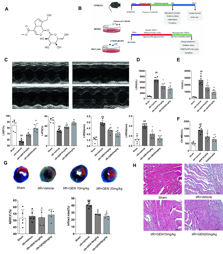
Methods: The MIRI model of animal was created in male C57BL/6J mice and the hypoxia reoxygenation (H/R) model was established for the in vitro experiments. Neonatal rat ventricular myocytes (NRVMs) and H9c2 cells with knockdown of TXNIP or NLRP3 were used. Geniposide was administered to mice before vascular ligation. HE staining, 2,3,5-triphenyltetrazolium chloride (TTC) staining, echocardiography, oxidative stress and myocardial enzyme detection were used to evaluate the cardioprotective effect of geniposide. Meanwhile, pharmacological approaches of agonist and inhibitor were used to observe potential pathway for geniposide cardioprotective in vitro and in vivo. Moreover, ELISA kits were adopted to detect the levels of inflammatory factors, such as IL-1β and IL-18. The gene and protein expression of NLRP3 and pyroptosis-related factors in heart tissue were performed by RT-PCR, western blotting and immunofluorescence in vivo and in vitro, respectively.

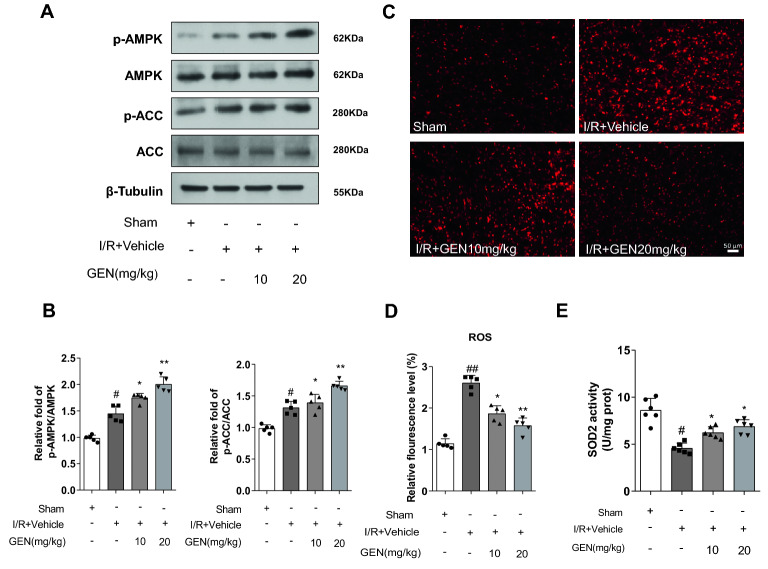
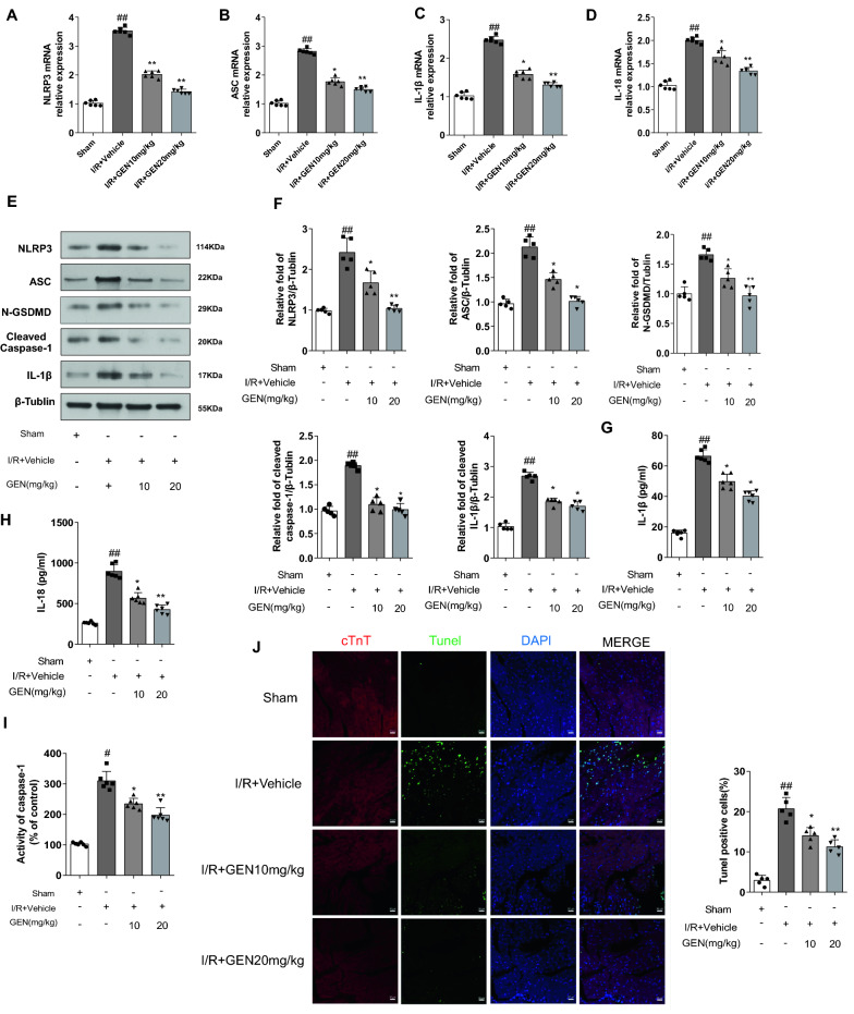
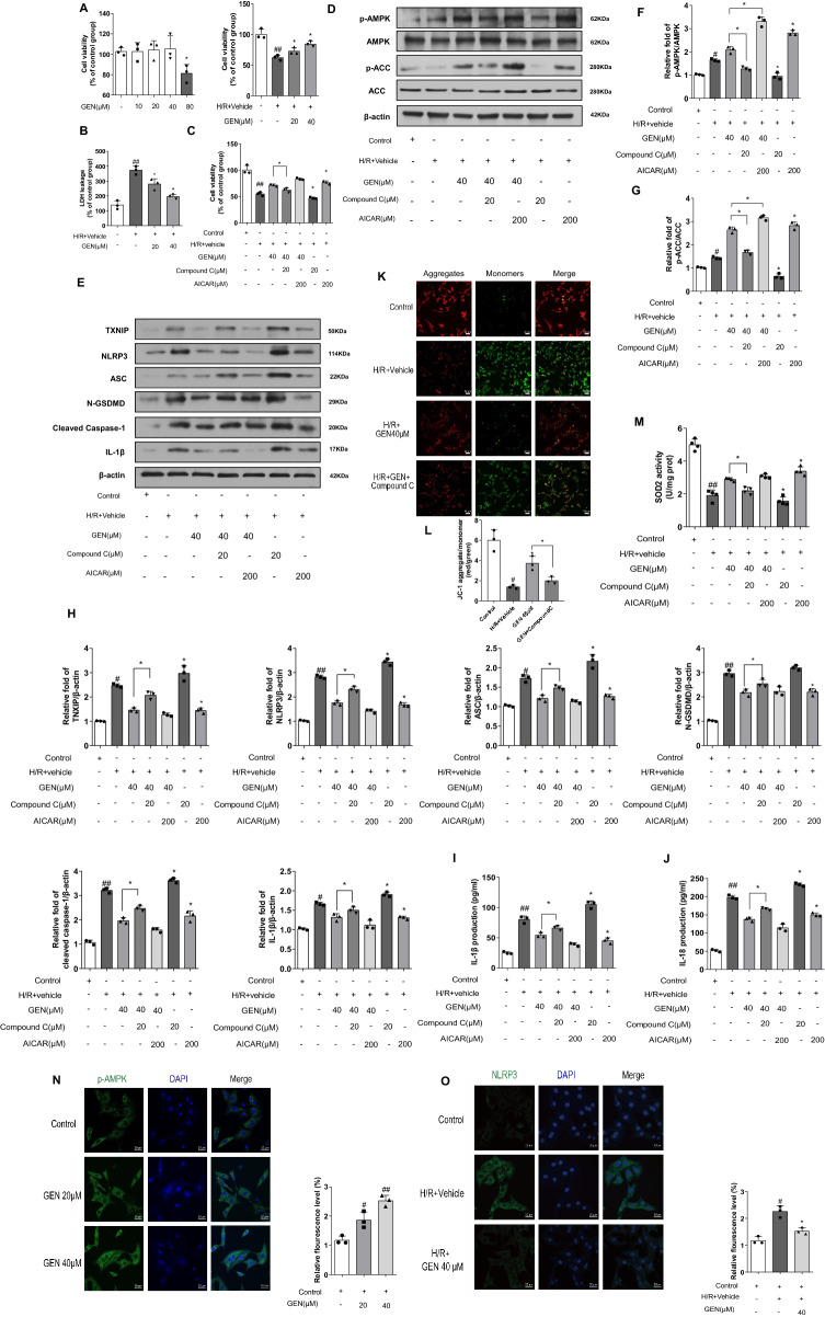
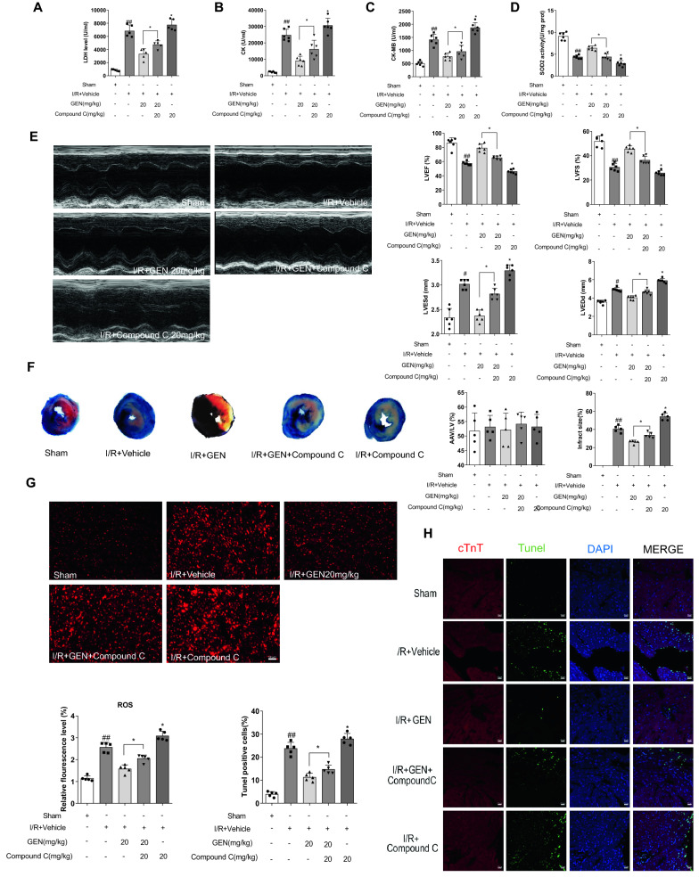
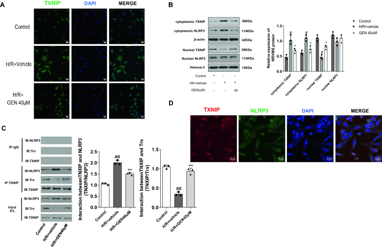
Results: Our results indicate that geniposide can reduce the area of myocardial infarction, improve heart function, and inhibit the inflammatory response in mice after MI/RI. In addition, RT-PCR and western blotting shown geniposide promoting AMPK phosphorylation to activate myocardium energy metabolism and reducing the levels of genes and proteins expression of NLRP3, ASC, N-GSDMD and cleaved caspase-1, IL-1β, IL-18. Meanwhile, geniposide improved NRVMs energy metabolism, which decreased ROS levels and the protein expression of TXNIP and thus suppressed the expression of NLRP3. AMPK antagonist or agonist and siRNA downregulation of TXNIP or NLRP3 were also verify the effect of geniposide against H/R injury. Further research found that geniposide promoted the translocation of TXNIP and reduce the binding of TXNIP and NLRP3.

Conclusions: In our study, geniposide can significantly inhibit NLRP3 inflammasome activation via the AMPK signaling pathway and inhibit pyroptosis of cardiomyocytes in myocardial tissues.

Keywords: AMPK; Geniposide; Myocardial ischemia reperfusion; NLRP3 inflammasome; Pyroptosis.


Conflict of interest statement

The authors have declared that no competing interest exists.

Figures

Fig. 1
Fig. 1
Geniposide improves cardiac function of mice suffering from MI/RI. A The chemical structure of geniposide. B The experimental design of our work. To examinate the effect of geniposide on MI/RI, the animals were divided into GEN treatment, I/R + Vehicle, and sham groups. To determine the mechanism of action of geniposide, another set of animals was administered with either Compound C or AICAR immediately at the beginning of reperfusion. The H/R model was launched in NRVMs and H9c2 cells. Compound C and AICAR, si-TXNIP, or si-NLRP3 were adopted to reveal the roles of pyroptosis and the AMPK/TXNIP/NLRP3 signaling on the cardioprotective effect of geniposide. C Representative echocardiograms in M-mode records of left ventricular (LV) and analysis of LV ejection fraction (LVEF), fractional shortening (LVFS), LV end-diastole diameter (LVEDd), and LV end-systolic diameter (LVESd) (n = 6). DF The activity of myocardial enzymes LDH, CK-MB, and CK levels in serum of mice (n = 6). G Representative TTC–Evans Blue stained sections of hearts and quantitative data of the LV infarct size. Infarct size (%) was expressed as the percentage of infarct area relative to the total left ventricular area. the nonischemic section shown in blue area, red represent risk area, and the infarct region is stained white (n = 6). H Representative H&E staining of the left ventricular area. Scale bar = 50 μm (n = 5). The data for each group were shown as the mean ± SD; #p < 0.05, ##p < 0.01 vs. Sham group; *p < 0.05, **p < 0.01 vs. I/R + Vehicle group
Fig. 2
Fig. 2
Geniposide alleviates cardiac damage caused by MI/R injury. A The protein expression of p-AMPK, AMPK, p-ACC, and ACC in mouse myocardium presented by representative western blot bands (n = 5). B The protein expression of p-AMPK and p-ACC in mice cardiac tissues of each group were shown by statistical histograms of representative western blots. C The representative DHE staining of the left ventricular of the mice, scale bar = 100 μm (n = 6). D Quantitative analyses of DHE staining level. E SOD2 activity of mouse heart samples were assayed using a commercial kit (n = 6). The data for each group were shown as the mean ± SD; #p < 0.05, ##p < 0.01 vs. Sham group; *p < 0.05, **p < 0.01 vs. I/R + Vehicle group
Fig. 3
Fig. 3
GEN exerts its protective effect by activating AMPKα and suppressing NLRP3 inflammasome activity. AD The mRNA expression of NLRP3, ASC, IL-1β, and IL-18 in mice myocardium of each group detected by RT-PCR (n = 5). E Representative western blotting results of NLRP3, ASC, N-GSDMD, cleaved caspase-1, and IL-1β (n = 6). F Densitometry data for NLRP3, ASC, N-GSDMD, cleaved caspase-1, and IL-1β in myocardium of each group of mice. G, H Inflammatory factor of IL-1β and IL-18 in the serum after GEN treatment (n = 5). I Myocardial caspase-1 activity was assessed in each group (n = 5). J Immunofluorescence TUNEL staining after GEN treatment (n = 6); scale bar = 20 μm. The data for each group were shown as the mean ± SD; #p < 0.05, ##p < 0.01 vs. Sham group; *p < 0.05, **p < 0.01 vs. I/R + Vehicle group
Fig. 4
Fig. 4
GEN ameliorates the cell injuries caused by H/R in NRVMs. A CCK-8 assay detected cell viability of NRVMs (n = 3). The results are presented as a percentage of the control. Cell viability of NRVMs following different concentrations of GEN exposure was measured by CCK-8 assay. NRVMs were treated with 20 μM and 40 μM with or without H/R insult. B, C Cell supernatants of LDH leakage and the change of cell viability in NRVMs. The cells were treated with the indicated treatments. D, E Expression of p-AMPK, AMPK, p-ACC, ACC, TXNIP, NLRP3, ASC, cleaved caspase-1, N-GSDMD-, IL-1β proteins detected by western blotting (n = 3). F, H Densitometry data of p-AMPK, p-ACC, ACC, TXNIP, NLRP3, ASC, cleaved caspase-1, N-GSDMD-, IL-1β in myocardium of each group of mice. I, J IL-1β and IL-18 in cell supernatants were detected by ELISA assays. K, L Intracellular red and green fluorescence of JC-1 was determined using a confocal microscope. The aggregates and monomers were assessed (n = 3); scale bar = 50 μm. M SOD2 enzymatic activity of NRVMs assayed with a commercial kit. N Representative images of immunofluorescence staining of p-AMPK in cardiomyocytes after different doses of GEN treated and the calculated intensity of p-AMPK (n = 3); scale bar = 20 μm. O Representative images of immunofluorescence staining of NLRP3 after H/R insult with 40 μM GEN treatment and the calculated intensity of NLRP3 (n = 3); scale bar = 20 μm. The data for each group were shown as the mean ± SD; #p < 0.05, ##p < 0.01 vs. Control group; *p < 0.05, **p < 0.01 vs. H/R + Vehicle group
Fig. 5
Fig. 5
Compound C counteracts the cardioprotective effects of geniposide in mice. AC Quantification of myocardium tissue (LDH, CK, CK-MB) on plasma of MI/RI mice treated with Compound C or GEN (n = 6). D SOD2 enzymatic activity of mice as indicated group assayed with a commercial kit (n = 5). E Quantitative assessment of analysis of LV ejection fraction (LVEF), fractional shortening (LVFS), left ventricular end-diastole diameter (LVEDd), and LV end-systolic diameter (LVESd) (n = 6). F Representative TTC–Evans Blue stained sections of hearts and quantitative data of the LV infarct size. Infarct size (%) was expressed as the percentage of infarct area relative to the total left ventricular area. the nonischemic section shown in blue area, red represent risk area, and the infarct region is stained white (n = 6). G Detection of ROS in myocardium by DHE staining. Compared with sham, the DHE fluorescence intensity in the myocardium of the vehicle-treated infarcted group was sharply increased and Compound C ablated the protective effect of geniposide (n = 6). H Immunofluorescence TUNEL staining after Compound C or GEN treatment (n = 6). The data for each group were shown as the mean ± SD; #p < 0.05, ##p < 0.01 vs. Sham group; *p < 0.05, **p < 0.01 vs. I/R + Vehicle group
Fig. 6
Fig. 6
Geniposide inhibits the activation of the TXNIP/NLRP3 complex formation in NRVMs. A, B Translocation of TXNIP occurs in NRVMs. Immunofluorescence staining for TXNIP, where DAPI is used to locate the nuclei of the cells, and Western blotting of TXNIP in the 3 groups (n = 3). TXNIP located in nucleus in control group and shifted to cytoplasm in H/R + Vehicle group, and geniposide at 40 μM reversed the TXNIP localization from the cytoplasm to nucleus. C After reoxygenation, the interaction of TXNIP/NLRP3 was determined by coimmunoprecipitation (Co-IP) in the NRVMS of the 3 groups. Cardiomyocytes samples of the H/R + Vehicle and GEN 40 μM groups were harvested after reoxygenation (n = 3). D Quantitative analysis of TXNIP/NLRP3. The data for each group were shown as the mean ± SD; #p < 0.05, ##p < 0.01 vs. Control group; *p < 0.05, **p < 0.01 vs. H/R + Vehicle group
Fig. 7
Fig. 7
Effect of TXNIP and NLRP3 siRNA on inflammasome of H/R stimulated H9c2 cells. After application of negative control siRNA (NC siRNA) or TXNIP and NLRP3 siRNA for 48 h, H9c2 cells were exposed to hypoxia for another 3 h, and subsequently reoxygenation for 2 h. AC Protein expression of TXNIP and NLRP3 was detected by Western blot at 48 h post-transfection, mRNA levels of TXNIP and NLRP3 in H9c2s were detected by RT-PCR at 24 h post-transfection and protein expression levels of ASC, N-GSDMD, cleaved Caspase-1 and IL-1β were analyzed by western blot. D, E Cell viability and LDH leakage after GEN treatment or using si-TXNIP or siNLRP3 (n = 3). F SOD2 enzymatic activity of H9c2 cells for the indicated groups, assayed using a commercial kit(n = 3). G IL-1β and IL-18 in cell supernatants were determined by ELISA assays. The data are presented as mean ± SD from three independent experiments. The data for each group were shown as the mean ± SD; #p < 0.05, ##p < 0.01 vs. Control group; *p < 0.05, **p < 0.01 vs. H/R + Vehicle group
Fig. 8
Fig. 8
The underlying molecular mechanisms of the protective effects of geniposide against the myocardial ischemia/reperfusion injury of mice. Scheme summarizing the protective effects of GEN on MI/RI via activating of AMPK signaling and inhibiting of ROS/TXNIP/NLRP3 mediating inflammation and subsequent pyroptosis-related signaling pathways. Under physiological conditions, Trx binds to TXNIP in its reduced form and suppresses activity. However, MI/RI amplifies ROS overproduction, which results in Trx dissociating from TXNIP, and thereby TXNIP/NLRP3 activation. This process leads to inflammation and pyroptosis. Importantly, GEN upregulates the AMPK signaling pathway, contributing to inhibit MI/RI-induced oxidative stress and TXNIP/NLRP3 activation, which were effectively inhibition inflammation and subsequent pyroptosis

References

    1. Zeng H, Wang L, Zhang J, Pan T, Yu Y, Lu J, et al. βActivated PKB/GSK-3 synergizes with PKC-signaling in attenuating myocardial ischemia/reperfusion injury potentiation of NRF2 activity: therapeutic efficacy of dihydrotanshinone-I. Acta Pharm Sin B. 2021;11(1):71–88. doi: 10.1016/j.apsb.2020.09.006. - DOI - PMC - PubMed
    1. Carling D. AMPK signalling in health and disease. Curr Opin Cell Biol. 2017;45:31–37. doi: 10.1016/j.ceb.2017.01.005. - DOI - PubMed
    1. Kahn B, Alquier T, Carling D, Hardie D. AMP-activated protein kinase: ancient energy gauge provides clues to modern understanding of metabolism. Cell Metab. 2005;1(1):15–25. doi: 10.1016/j.cmet.2004.12.003. - DOI - PubMed
    1. Wu S, Zou M. AMPK, mitochondrial function, and cardiovascular disease. Int J Mol Sci. 2020;21(14):4987. doi: 10.3390/ijms21144987. - DOI - PMC - PubMed
    1. Dolinsky V, Chan A, Robillard Frayne I, Light P, Des Rosiers C, Dyck J. Resveratrol prevents the prohypertrophic effects of oxidative stress on LKB1. Circulation. 2009;119(12):1643–1652. doi: 10.1161/CIRCULATIONAHA.108.787440. - DOI - PubMed

LinkOut - more resources