Review
. 2022 Jun 17;13(1):257.
doi: 10.1186/s13287-022-02944-7.

Spotlight on therapeutic efficiency of mesenchymal stem cells in viral infections with a focus on COVID-19

Affiliations
Review

Spotlight on therapeutic efficiency of mesenchymal stem cells in viral infections with a focus on COVID-19

Saman Yasamineh et al. Stem Cell Res Ther. .

Abstract

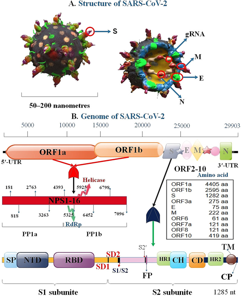
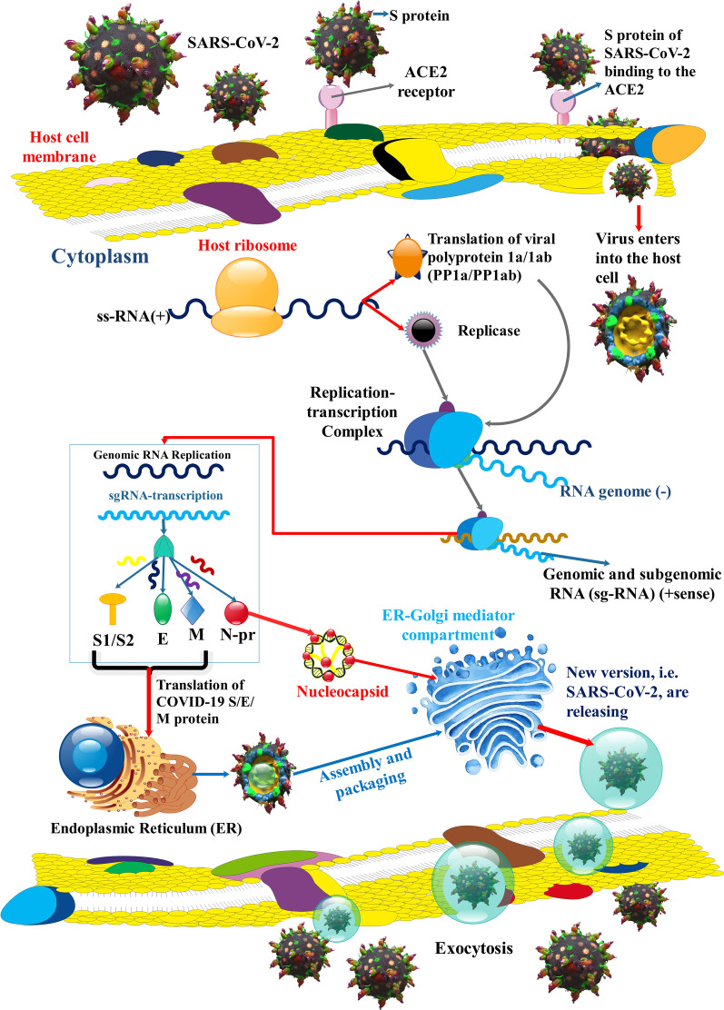
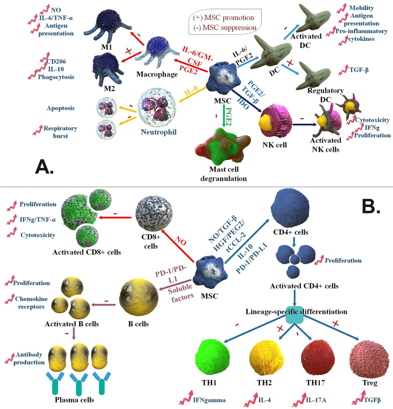
The SARS-COV-2 virus has infected the world at a very high rate by causing COVID-19 disease. Nearly 507 million individuals have been infected with this virus, with approximately 1.2% of these patients being dead, indicating that this virus has been out of control in many countries. While researchers are investigating how to develop efficient drugs and vaccines versus the COVID-19 pandemic, new superseded treatments have the potential to reduce mortality. The recent application of mesenchymal stem cells (MSCs) in a subgroup of COVID-19 patients with acute respiratory distress has created potential benefits as supportive therapy for this viral contagion in patients with acute conditions and aged patients with severe pneumonia. Consequently, within this overview, we discuss the role and therapeutic potential of MSCs and the challenges ahead in using them to treat viral infections, with highlighting on COVID-19 infection.

Keywords: COVID-19; Mesenchymal stem cells; SARS-CoV-2; Stem cells therapy.


Conflict of interest statement

The authors have no relevant financial or non-financial interests to disclose.

Figures

Fig. 1
Fig. 1
3D-graphical illustrations of the structural proteins of SARS-CoV-2 (A) and genome (B). S protein genetic site contained SP (single peptide), CP (cytoplasmic tail), FP (fusion peptide), CD (connector domain), NTD (N-terminal domain), RBD, TM (transmembrane domain), SD1, and SD2 (subdomain 1 and 2), S1/S2 (S1/S2 protease cleavage site), S2′ (S2′ protease cleavage site), HR1, CH (central helix), HR2
Fig. 2
Fig. 2
SARS-CoV-2 replication. SARS-CoV-2 S protein connects to ACE2 receptor to membrane fusion and discharges the viral genome into the host cell cytoplasm. After the entering the virus, the viral RNA is uncovered in the cytoplasm. ORF1a and ORF1ab are translated to produce pp1a and pp1ab that undergo further cleavage into smaller proteins containing RNA-dependent RNA polymerase, helicase, and nonstructural protein. RNA replicase–transcriptase complex (RTC) localizes to altered intracellular membranes isolated from the rough endoplasmic reticulum (ER) in the perinuclear area, where it generates (−) RNAs. Throughout replication, complete (−)RNA transcripts of the genome are generated and used as patterns for full (+) RNA genomes. Throughout transcription, a member of subgenomic RNAs (sg-RNA), containing those encoding S, M, E, and E protein, is generated by intermittent transcription. In this procedure, a nested set of (−)sg-RNA is generated that modify in length at the 3′ end and 5′-leader sequence, which is essential for translation. Then, this (−) sg-RNA are transcribed into (+) sg-mRNAs. Afterward, S, M, E, and N proteins are gathered in the nucleocapsid and viral envelope at the ER–Golgi intermediate compartment (ERGIC), then through exocytosis of SARS-CoV-2, released to vesicles [–131]
Fig. 3
Fig. 3
Possible immune system reactions in COVID-19 patients. To create an antiviral response, the innate immune system requires to diagnose the invasion of the viral, frequently via PAMPs. For the SARS-CoV-2, it is recognized that PAMPs in the shape of viral genomic RNA or the mediators in viral replication, such as dsRNA, are identified via either the endosomal RNA receptors, TLR3 and TLR7 and the cytosolic RNA sensor, and retinoic acid-inducible gene I/melanoma differentiation-associated gene 5 (RIG-I/MDA5). This detection occurrence triggers the downstream signaling cascade, including IRF3 and NF-κB, accompanied by their nuclear translocation. Type I IFN by interferon-α/β receptor (IFNAR), respectively, triggers the JAKs, STATs pathway, where JAK1 and tyrosine kinase 2 (TYK2) kinases phosphorylate STAT1 and STAT2. STAT1/2 organize a complex by IRF9, and simultaneously they transfer to the nucleus to initiate the transcription of IFN-stimulated genes (ISGs) with the regulation of IFN-stimulated response element (ISRE) comprising promoters [51]. Moreover, the SARS-CoV-2 is possible that trigger the inflammasome sensor, NACHT, LRR, and PYD domains-containing protein 3 (NALP3), leading to the release of the greatly inflammatory cytokine IL-1β and cause of pyroptosis, which is an inflammatory led to cell death. Envelope proteins and 3a-nsp of SARS caused the creation of the NLRP3 inflammasome [132, 133]
Fig. 4
Fig. 4
MSCs function as immunosuppression of the adaptive and innate immune system. A MSCs use various molecular pathways to inhibit innate immune cells. MSCs inhibit macrophage polarization to M1 via favors M2 polarization. MSCs suppress mast cell degranulation of histamine-comprising granules and suppress DC and NK cell activation, differentiation, and effector actions. MSC-isolated PGE2 chips in the whole of this efficacy. MSC-generated IL-6 inhibits neutrophil apoptosis and respiratory burst and helps suppress DC action. In the presence of GM-CSF and IL-6, MSCs also affect macrophage action, while IDO and TGF-β inhibit NK cell action. Moreover, MSCs favor the production of regulatory DCs. B MSCs suppress different facets of B cells acting, such as activation, reproduction, chemokine receptor expression, and differentiation to changing antibody-releasing plasma cells. Unknown soluble factors and programmed death-1 (PD-1)/PD ligand-1 (PD-L1) ligation intercede the efficacy of MSCs on B cells. MSC induced NO in reaction to inflammatory cytokine diagnosis to inhibit CD8+ T cell proliferation, cytokine generation, and cytotoxicity. In reaction to activation in a particular cytokine milieu, CD4+ T cells can differentiate into many effector crowds. MSCs generate soluble factors (IL-10, truncated CCL-2, PGE2, HGF, TGF-β, and NO) and membrane-bound molecules (PD-1 ligation) to inhibit Th cell reproduction and the polarization of CD4+ T cells to TH17 and TH1 cells. MSCs favor the growth of anti-inflammatory Treg and TH2 crowds [134]
Fig. 5
Fig. 5
Different functions [Inhibition (I: red arrows) and Activation (A: blue arrows)] of MSC-EVs in various tissues injury. BM-MSCs Exos reduced macrophage influx, repressed inflammatory intermediaries, including MCP-1, and suppressed STAT3 signaling in the lung injury. Besides, UC-MSC EVs recovered lung functions by bronchopulmonary dysplasia through increasing M1-M2 change in macrophages showed by decreased IL-6, TNF-α, CCL5, and enhanced arginase 1. BM-MSC EVs diminished bowel injury, limited myeloperoxidase action in the colon, reduced IL-1β, TNF-α, and inducible nitric oxide synthase (iNOS) amounts in the bowel. Moreover, upon UC-MSC EVs therapy, macrophage penetration to the tissue was decreased along with downregulation of TNF-α, IL-1β, IL-6, IL-7, and iNOS in colon tissue and spleen. BM-MSC EVs are protected in a murine renal I/R injury model and the preservative efficacy was associated with the C–C chemokine receptor type 2 transported on the exosome surface, which could separate the chemokine (C–C motif) ligand 2 (CCL2) and thus damaged macrophage recruitment and activation. Moreover, UC-MSC EVs decreased the amounts of IL-1β and TNFα while increasing autophagy. Human embryonic stem cell-isolated MSCs decreased oxidative stress, enhanced myocardial livability via the triggering of the PI3K/Akt pathway, and therefore increased cardiac activity. Besides, BM-MSC EVs enhanced angiogenesis and increased blood circulation in myocardial infarction models while preventing T cell proliferation. HUC-MSCs EVs were displayed to decrease hepatic inflammation while reducing TGF-β amounts and collagen deposition in murine liver fibrosis. Moreover, BM-MSC EVs infusion decreases IFN-γ, IL-1, IL-2, TNF-α expression, enhances Treg level, and reduces necrosis in the liver. MSC-EVs in rheumatoid arthritis suppress T cell proliferation and also promotion of Treg cells, Breg cells [110]
Fig. 6
Fig. 6
COVID-19 leads to cell and damage injury, discharge of danger-associated molecular patterns (DAMPs), PAMPs, and inflammatory intermediates that increase immune cell penetration. Injected MSCs-EVs can decrease inflammation and induce tissue regeneration. Significantly, the good efficacy of MSCs is displayed to be related to the decrease in TNF-α, interleukin-1/6 via the discharge of HGF, PGE2, lipoxin A4 (LXA4), and TSG-6, inhibition of inflammatory T cell proliferation via indoleamine 2,3-dioxygenase expression, switch from Th1 and Th17 reactions to Th2, and suppression of monocytes and myeloid DCs maturation. MSCs stimulate M2 polarization via juxtacrine signaling and paracrine agents, including HGF, PGE2, and TSG-6, leading to a monocyte helping enhance anti-inflammatory IL-10, which synergistically induces Treg cells and activates tissue regeneration pathways. Additionally, MSC involvement in several mechanisms that increase lung fibrosis leads to protecting efficacy, as shown in various lung damage models. Notably, MSCs decrease the damage-associated alveolar edema and endothelial permeance via the discharge of keratinocyte KGF to increase the sodium-affiliate alveolar fluid clearance via epithelial sodium channel (ENaC) and have anti-apoptotic and anti-oxidative pathways to repair cytokine-injured alveolar type II cells and epithelial and endothelial damage via the discharge of angiopoietin-1, lipoxin A4 (LXA4) and TSG-6 [135]

Similar articles

Cited by

References

    1. Hashemi B, et al. Emerging importance of nanotechnology-based approaches to control the COVID-19 pandemic; focus on nanomedicine iterance in diagnosis and treatment of COVID-19 patients. J Drug Deliv Sci Technol. 2021;144:102967. - PMC - PubMed
    1. Taghavi-farahabadi M, et al. Hypothesis for the management and treatment of the COVID-19-induced acute respiratory distress syndrome and lung injury using mesenchymal stem cell-derived exosomes. Med Hypotheses. 2020;144:109865. - PMC - PubMed
    1. Long Q-X, et al. Antibody responses to SARS-CoV-2 in patients with COVID-19. Nat Med. 2020;26:845–848. - PubMed
    1. Coronavirus N. Situation reports-World Health Organization (WHO). 2019.
    1. Khailany RA, Safdar M, Ozaslan M. Genomic characterization of a novel SARS-CoV-2. Gene Rep. 2020;19:100682. - PMC - PubMed