Review
. 2022 Sep;28(9):710-725.
doi: 10.1016/j.molmed.2022.05.010. Epub 2022 Jun 15.

Can't handle the stress? Mechanobiology and disease

Affiliations
Review

Can't handle the stress? Mechanobiology and disease

Noam Zuela-Sopilniak et al. Trends Mol Med. 2022 Sep.

Abstract

Mechanobiology is a rapidly growing research area focused on how mechanical forces and properties influence biological systems at the cell, molecular, and tissue level, and how those biological systems, in turn, control mechanical parameters. Recently, it has become apparent that disrupted mechanobiology has a significant role in many diseases, from cardiovascular disease to muscular dystrophy and cancer. An improved understanding of this intricate process could be harnessed toward developing alternative and more targeted treatment strategies, and to advance the fields of regenerative and personalized medicine. Modulating the mechanical properties of the cellular microenvironment has already been used successfully to boost antitumor immune responses and to induce cardiac and spinal regeneration, providing inspiration for further research in this area.

Keywords: Mechanobiology; extracellular matrix; human disease; mechanotransduction; microenvironment; tissue mechanics.


Conflict of interest statement

Declaration of interests None declared by authors.

Figures
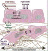
Figure 1:
Figure 1:. Cells in tissues are exposed to numerous sources of mechanical forces that are sensed by the cells and that modulate the cellular fate and function.
(i) Fluid shear stress resulting from blood and lymph flow through the circulatory and lymphatic systems, as well as interstitial fluid flow. (ii) Hydrostatic pressure from the circulatory and lymphatic systems and/or interstitial fluid. The more fluid that filters into the interstitial space from the vasculature, the greater the hydrostatic pressure within the tissue. (iii) Cell-cell interactions, such as pushing and/or pulling forces from neighboring cells, or during collective migration or interaction with other cells such as tumor associated macrophages or during immune cell activation. (iv) Cells can sense differences in the stiffness, structure, and composition of the extracellular matrix via cell surface receptors such as integrins and their cytoplasmic connections. Extracellular matrix stiffness is frequently altered in pathological conditions such as tumorigenesis or fibrosis, modulating cellular functions. (v) Cells can experience physical confinement when migrating through small openings in the extracellular matrix or tight interstitial spaces, or from the presence of neighboring cells. (vi) Traction forces can be exerted from migrating cells to the extracellular matrix. (vii) Large scale tissue deformation, for example contraction of muscle, stretching of skin, or compressive loads applied to cartilage and bone, result in tensile, compressive, and/or shear forces on the cells within the tissue.
Figure 2:
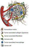
Figure 2:. The tumor microenvironment.
Tumor cells manipulate both cellular and non-cellular elements in their microenvironment to thrive under hostile conditions and facilitate tumor progression. The resulting tumor microenvironment (TME) consists mainly of hematopoietic immune cells such as T cells, B cells, natural killer cells, dendritic cells and tumor associated macrophages (TAMs) that in collaboration with resident stromal cells such as cancer associated fibroblasts (CAFs) play a key role in tumorigenesis and tumor progression through matrix remodeling and generation of specialized tumor ECM structures such as tumor associated collagen signatures (TACS). The extracellular matrix serves as a scaffold and represents the non-cellular component of the TME. The different elements of the TME interact through the different ECM components, cell-cell contacts, and the release of cytokines and chemokines, among others.
Figure 3:
Figure 3:. Mechanobiology of T cell activation.
The interaction of T cells with antigen presenting cells (APC) is a crucial step of the adaptive immune response. Discrete cytoplasmic actin structures apply forces at different areas of the T cell–APC interaction, facilitating T cell activation. (i) Upon initial contact between a T cell and an APC, protrusive actin structures push into the APC to overcome the glycocalyx and create areas of close contact between the T cell and APC cell membranes. Subsequent retraction of these dynamic structures creates tension on the TCR–pMHC bonds that facilitates antigen discrimination between self and cognate pMHCs and TCR activation, as only strong, highly specific TCR– cognate pMHC bonds will withstand the applied forces. (ii) At the leading edge of the migrating T cell, actin-driven lamellipodial protrusions apply force on the interacting APC, allowing for TCR triggering and signal accumulation. This region contains a prominent branched actin network generated by the Arp2/3 complex activator WAVE2. The same mechanism is in play in early stages of the formation of a stable immune synapse, where initial synapse triggering induces spreading of the T cell on the APC surface. (iii) Myosin contractility drives retrograde flow of actin bundles that bend as they move inward and are cross-linked by myosin IIA. The resulting actomyosin arcs sweep TCR MHCs toward the center of the cell, promoting sequential triggering of many TCR molecules by a single agonist pMHC.

References

    1. Agarwal P and Zaidel-Bar R, “Mechanosensing in embryogenesis,” Current Opinion in Cell Biology, vol. 68, pp. 1–9, 2021, doi: 10.1016/j.ceb.2020.08.007. - DOI - PubMed
    1. Ranade SS, Syeda R, and Patapoutian A, “Mechanically Activated Ion Channels,” Neuron, vol. 87, no. 6, pp. 1162–1179, 2015, doi: 10.1016/j.neuron.2015.08.032. - DOI - PMC - PubMed
    1. Maurer M and Lammerding J, “The Driving Force: Nuclear Mechanotransduction in Cellular Function, Fate, and Disease,” Annual Review of Biomedical Engineering, vol. 21, no. 1, pp. 443–468, Jun. 2019, doi: 10.1146/annurev-bioeng-060418-052139. - DOI - PMC - PubMed
    1. Massou S et al., “Cell stretching is amplified by active actin remodelling to deform and recruit proteins in mechanosensitive structures,” Nature Cell Biology, vol. 22, no. 8, pp. 1011–1023, 2020, doi: 10.1038/s41556-020-0548-2. - DOI - PubMed
    1. Alonso JL and Goldmann WH, “Cellular mechanotransduction,” AIMS Biophysics, vol. 3, no. 1, 2016, doi: 10.3934/biophy.2016.1.50. - DOI

Publication types