. 2022 Jun 20;18(1):233.
doi: 10.1186/s12917-022-03327-z.

Perioperative characteristics, histologic diagnosis, complications, and outcomes of dogs undergoing percutaneous drainage, sclerotherapy or surgical management of intrarenal cystic lesions: 18 dogs (2004-2021)

Affiliations

Perioperative characteristics, histologic diagnosis, complications, and outcomes of dogs undergoing percutaneous drainage, sclerotherapy or surgical management of intrarenal cystic lesions: 18 dogs (2004-2021)

Michail Vagias et al. BMC Vet Res. .

Abstract

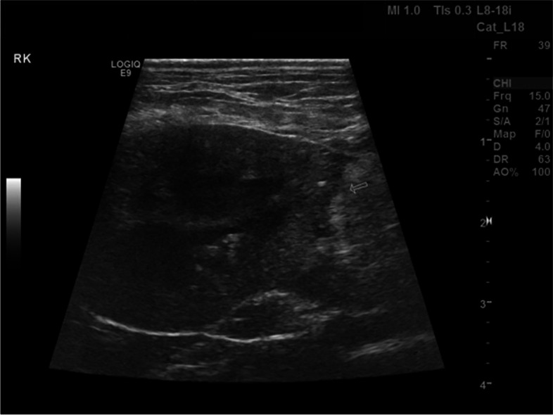
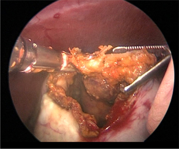
Background: Canine intrarenal cystic lesions (ICLs) are infrequently reported in the veterinary literature. Several treatment options have been described including cyst fenestration (partial nephrectomy/deroofing) +/- omentalization, sclerotherapy using alcohol as a sclerosing agent, percutaneous cyst drainage (PCD), and ureteronephrectomy. Information regarding presenting clinical signs, physical examination findings, histologic diagnosis and outcomes of dogs with ICLs treated by different methods is limited. Medical records of 11 institutions were retrospectively reviewed to identify dogs that underwent PCD, sclerotherapy, surgical deroofing +/- omentalization, or ureteronephrectomy for management of ICLs from 2004 to 2021. Six weeks postoperative/post-procedural follow-up was required. Cases suspected to represent malignancy on preoperative imaging were excluded. The study objective was to provide information regarding perioperative characteristics, complications, and outcomes of dogs undergoing treatment of ICLs.

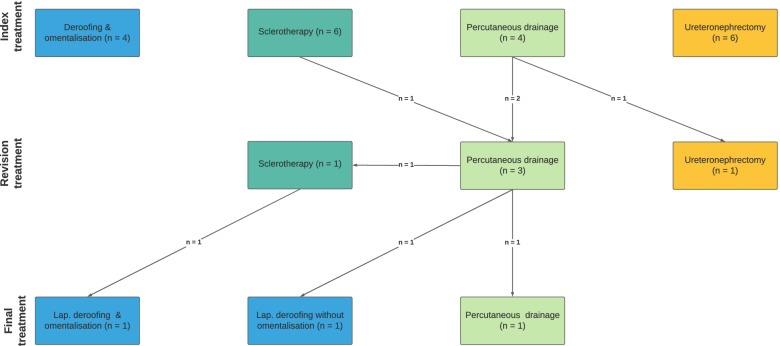
Results: Eighteen dogs were included, with 24 ICLs treated. Ten had bilateral. There were 15 males and 3 females, with crossbreeds predominating. PCD, sclerotherapy, deroofing and ureteronephrectomy were performed in 5 (5 ICLs treated), 7 (11 ICLs), 6 (6), and 7 (7) dogs, respectively, with 5 dogs undergoing > 1 treatment. Seven dogs experienced 8 complications, with requirement for additional intervention commonest. PCD, sclerotherapy and deroofing resulted in ICL resolution in 0/5, 3/11 and 3/6 treated ICLs, respectively. Histopathology identified renal cysts (RCs) in 7/13 dogs with histopathology available and neoplasia in 6/13 (4 malignant, 2 benign). Of 5 dogs diagnosed histopathologically with neoplasia, cytology of cystic fluid failed to identify neoplastic cells. Among 7 dogs with histologically confirmed RCs, 4 had concurrent ICLs in ipsilateral/contralateral kidney, compared with 2/6 dogs with histologically confirmed neoplasia.

Conclusions: Benign and neoplastic ICLs were approximately equally common and cystic fluid cytology failed to differentiate the 2. Among renal-sparing treatments, deroofing most commonly resulted in ICL resolution. Presence of concurrent ICLs in ipsilateral/contralateral kidney does not appear reliable in differentiating benign from malignant ICLs.

Keywords: Cyst drainage; Deroofing; Dog; Intrarenal cystic lesion; Renal cyst; Sclerotherapy.


Conflict of interest statement

The authors declare that they have no competing interests.

Figures

Fig. 1
Fig. 1
Lateral (a) and ventrodorsal (b) abdominal radiographs of a 5.2-year-old, 16.5 kg, neutered male, mixed breed dog treated with deroofing and diagnosed with renal cyst. A large ovoid soft tissue opacity is identified within the right craniodorsal abdomen, which causes a significant mass effect on surrounding abdominal structures. Images courtesy of Dr. Mullins
Fig. 2
Fig. 2
Transverse (a), sagittal (b) and dorsal (c) plain computed tomographic multiplanar reconstruction images of the same dog diagnosed with renal cyst in Fig. 1. Multiple, non-contrast enhancing, well-defined, ovoid structures of variable size and of fluid attenuation are visible within the cortex and medulla of both kidneys. The caudal pole of the right kidney contains a very large, ovoid, thin-walled, non-contrasting enhancing structure of fluid attenuation that has replaced the normal architecture of its caudal third. For figures (a) and (c), the left side of the dog is at the right side of the images. Images courtesy of Dr. Mullins
Fig. 3
Fig. 3
Ultrasonographic long-axis image of the left kidney of a 8.8-year-old, 41.9 kg, neutered male, mixed breed dog diagnosed with ICL treated with sclerotherapy (histopathology not available). A large anechoic cystic structure is identified within the caudal pole of the left kidney effacing the middle third of the renal parenchyma, has rounded turgid walls and lacks septations. Cranial is to the right of the image. Image courtesy of Dr. Grimes
Fig. 4
Fig. 4
Flow diagram summarizing the different treatment procedures each dog underwent. Index treatment is defined as the first treatment performed
Fig. 5
Fig. 5
Ventrodorsal fluoroscopic image of a 14-year-old, 6.4 kg, neutered male, miniature pinscher diagnosed with renal cystadenocarcinoma of the right kidney. Intravenous contrast was injected percutaneously through a pigtail catheter to confirm its location and that the entirety of contrast remains within the ICL with no extracapsular leakage. Image courtesy of Dr. Flanders
Fig. 6
Fig. 6
Intraoperative image during deroofing of the renal cyst of the same dog in Fig. 1. The wall of the renal cyst has been incised and its inner aspect is presented with the use of Allis tissue forceps prior to completion of deroofing. Cranial is to the top left of the image. Image courtesy of Dr. Mullins
Fig. 7
Fig. 7
Intraoperative image during omentalization of the renal cyst of the same dog in Fig. 1 after deroofing. A double layer of a portion of the greater omentum in the region of the right kidney was sutured around the entire circumference of the remaining rim of cystic lining using absorbable monofilament suture in a simple continuous pattern. Cranial is to the right of the image. Image courtesy of Dr. Mullins
Fig. 8
Fig. 8
Long-axis ultrasonographic image of the right kidney of a 13-year-old, 5.6 kg, spayed female, Shih Tzu diagnosed with renal cyst of the right kidney. Image was obtained 6 weeks following deroofing and omentalization, and demonstrates the truncated appearance of the caudal pole of the kidney at the site of omentalization (arrow). Cranial is to the left of the image. Image courtesy of Dr. Mullins
Fig. 9
Fig. 9
Intraoperative image during laparoscopic deroofing of the renal cystadenocarcinoma of the same dog in Fig. 5. The cystic wall was partially resected using bipolar vessel sealing device. Image courtesy of Dr. Flanders
Fig. 10
Fig. 10
Macroscopic appearance of the excised left kidney (incised sagittally) of a 11.4-year-old, 29.1 kg, neutered male, giant poodle diagnosed with renal cyst. A thin-walled cyst is identified at caudal pole of left kidney replacing the normal parenchyma. Image courtesy of Dr. de Rooster

References

    1. Park J, Moon C, Lee M, Lee HB, Jeong SM. Surgical fenestration combined with omentalization for the treatment of renal cysts in 2 dogs. Can Vet J. 2019;60(10):1104–10. - PMC - PubMed
    1. Zatelli A, D'Ippolito P, Bonfanti U, Zini E. Ultrasound-assisted drainage and alcoholization of hepatic and renal cysts: 22 cases. J Am Anim Hosp Assoc. 2007;43(2):112–116. doi: 10.5326/0430112. - DOI - PubMed
    1. Agut A, Soler M, Laredo FG, Pallares FJ, Seva JI. Imaging diagnosis-ultrasound-guided ethanol sclerotherapy for a simple renal cyst. Vet Radiol Ultrasound. 2008;49(1):65–67. doi: 10.1111/j.1740-8261.2007.00319.x. - DOI - PubMed
    1. Orioles M, Di Bella A, Merlo M, Ter Haar G. Ascites resulting from a ruptured perinephric pseudocyst associated with a renal cyst in a dog. Vet Rec Case Rep. 2014;2:e000012.
    1. Paskalev M, Lazarov L, Atanasov A. A case of solitary renal cyst in a dog. Bulgarian J Vet Med. 2012;15(1):62–67.

LinkOut - more resources