

Post-synaptic specialization of the neuromuscular junction: junctional folds formation, function, and disorders
- PMID: 35718785
- PMCID: PMC9208267
- DOI: 10.1186/s13578-022-00829-z
Post-synaptic specialization of the neuromuscular junction: junctional folds formation, function, and disorders
Abstract
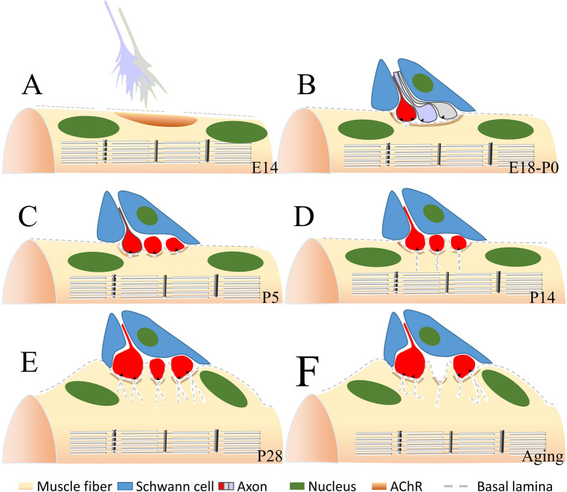
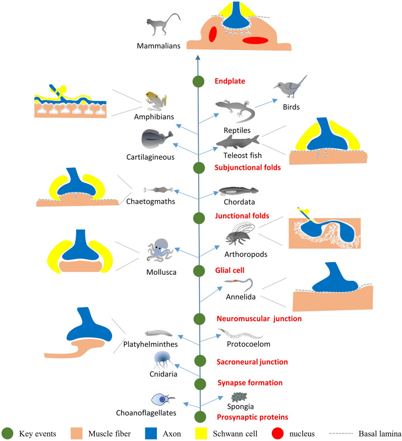
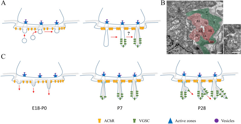
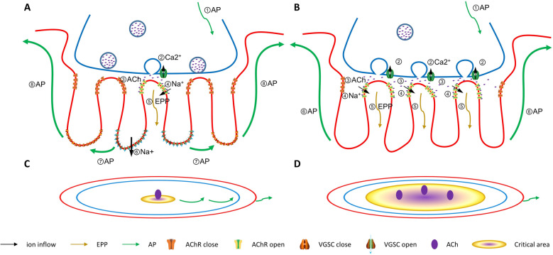
Post-synaptic specialization is critical to the neurotransmitter release and action potential conduction. The neuromuscular junctions (NMJs) are the synapses between the motor neurons and muscle cells and have a more specialized post-synaptic membrane than synapses in the central nervous system (CNS). The sarcolemma within NMJ folded to form some invagination portions called junctional folds (JFs), and they have important roles in maintaining the post-synaptic membrane structure. The NMJ formation and the acetylcholine receptor (AChR) clustering signal pathway have been extensively studied and reviewed. Although it has been suggested that JFs are related to maintaining the safety factor of neurotransmitter release, the formation mechanism and function of JFs are still unclear. This review will focus on the JFs about evolution, formation, function, and disorders. Anticipate understanding of where they are coming from and where we will study in the future.
Keywords: Development; Disease; Formation mechanism; Function; Junctional folds; Neuromuscular junction; Post-synaptic specialization.
© 2022. The Author(s).
Conflict of interest statement
The authors declare that they have no competing interests.
Figures
Similar articles
-
Abnormalities in neuromuscular junction structure and skeletal muscle function in mice lacking the P2X2 nucleotide receptor.Neuroscience. 2007 Sep 7;148(3):700-11. doi: 10.1016/j.neuroscience.2007.06.050. Epub 2007 Jul 17. Neuroscience. 2007. PMID: 17706883
-
Super-Resolution Microscopy Reveals a Nanoscale Organization of Acetylcholine Receptors for Trans-Synaptic Alignment at Neuromuscular Synapses.eNeuro. 2017 Aug 10;4(4):ENEURO.0232-17.2017. doi: 10.1523/ENEURO.0232-17.2017. eCollection 2017 Jul-Aug. eNeuro. 2017. PMID: 28798955 Free PMC article.
-
Overexpression of Dok-7 in skeletal muscle enhances neuromuscular transmission with structural alterations of neuromuscular junctions: Implications in robustness of neuromuscular transmission.Biochem Biophys Res Commun. 2020 Feb 26;523(1):214-219. doi: 10.1016/j.bbrc.2019.12.011. Epub 2019 Dec 14. Biochem Biophys Res Commun. 2020. PMID: 31848047
-
Dissecting the Extracellular Complexity of Neuromuscular Junction Organizers.Front Mol Biosci. 2020 Jan 10;6:156. doi: 10.3389/fmolb.2019.00156. eCollection 2019. Front Mol Biosci. 2020. PMID: 31998752 Free PMC article. Review.
-
Clustering of nicotinic acetylcholine receptors: from the neuromuscular junction to interneuronal synapses.Mol Neurobiol. 2002 Feb;25(1):79-112. doi: 10.1385/MN:25:1:079. Mol Neurobiol. 2002. PMID: 11890459 Review.
Cited by
-
Unraveling the causes of sarcopenia: Roles of neuromuscular junction impairment and mitochondrial dysfunction.Physiol Rep. 2024 Jan;12(1):e15917. doi: 10.14814/phy2.15917. Physiol Rep. 2024. PMID: 38225199 Free PMC article. Review.
-
Recognising the potential of large animals for modelling neuromuscular junction physiology and disease.J Anat. 2022 Nov;241(5):1120-1132. doi: 10.1111/joa.13749. Epub 2022 Sep 2. J Anat. 2022. PMID: 36056593 Free PMC article. Review.
-
Sarcoglycans are enriched at the neuromuscular junction in a nerve-dependent manner.Cell Death Dis. 2025 Jan 22;16(1):37. doi: 10.1038/s41419-025-07353-1. Cell Death Dis. 2025. PMID: 39843456 Free PMC article.
-
Nerve-independent formation of membrane infoldings at topologically complex postsynaptic apparatus by caveolin-3.Sci Adv. 2023 Jun 16;9(24):eadg0183. doi: 10.1126/sciadv.adg0183. Epub 2023 Jun 16. Sci Adv. 2023. PMID: 37327338 Free PMC article.
-
The molecular athlete: exercise physiology from mechanisms to medals.Physiol Rev. 2023 Jul 1;103(3):1693-1787. doi: 10.1152/physrev.00017.2022. Epub 2023 Jan 5. Physiol Rev. 2023. PMID: 36603158 Free PMC article. Review.
References
-
- Fox MA. Development of the vertebrate neuromuscular junction. In: Hortsch M, Umemori H, editors. The Sticky Synapse. Berlin: Springer; 2009.
Publication types
Grants and funding
LinkOut - more resources
Full Text Sources
