

Clue Cells and Pseudo Clue Cells in Different Morphotypes of Bacterial Vaginosis
- PMID: 35719334
- PMCID: PMC9198243
- DOI: 10.3389/fcimb.2022.905739
Clue Cells and Pseudo Clue Cells in Different Morphotypes of Bacterial Vaginosis
Abstract
Introduction: Clue cells (epithelial cells heavily covered with adherent bacteria) are an accepted clue to the diagnosis of bacterial vaginosis. However, the exact morphologic criteria of clue cells and bacterial adherence were never elaborated.
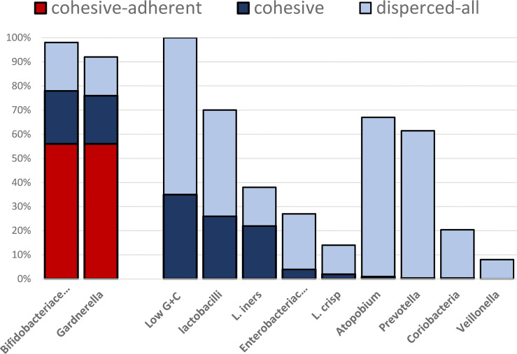
Materials and methods: We investigated adhesive and cohesive patterns of main microbiota groups in vaginal discharge using fluorescence in situ hybridization (FISH). Samples from 500 women diagnosed with bacterial vaginosis and positive for clue cells with classic microscopy were collected from 42 gynecologic practices in Berlin and reexamined in our FISH laboratory for the spatial distribution of Bifidobacteriaceae, Gardnerella, Fannyhessea vaginae (Atopobium); low G+C (guanine+cytosine) bacteria, lactobacilli, Lactobacillus iners; Lactobacillus crispatus, Gamma-Proteobacteria; and Enterobacteriaceae, Prevotella-Bacteroides, Veillonella, and Coriobacterium groups.
Results: Bacterial taxa present in vaginal smears were not accidentally assembled according to their relative abundance but were built in group-specific distribution patterns, which can be well described by two features: cohesiveness to each other and adherence to epithelial cells. Accordingly, four patterns can be distinguished: dispersed (non-adherent bacteria), dispersed adherent bacteria, cohesive (non-adherent) bacteria, and cohesive adherent bacteria. Direct cohesive adherence to the epithelial cells representing true clue cells was unique for Gardnerella species and observed only in 56% of the investigated samples. In the remaining vaginal samples, the epithelial cells were mechanically entrapped in bacterial masses, and the composition was unrelated to the epithelial cell surface, building non-adherent pseudo clue cells. The proportion of women with true clue cells in their samples from different gynecologic practices varied from 19% to 80%.
Discussion: Taxon indifferent imaging is inadequate for the exact analysis of the microbial layer adjacent to the vaginal epithelial cells. Morphologically seen bacterial vaginosis is a mix of at least two different conditions: biofilm vaginosis and bacterial excess vaginosis.
Keywords: FISH; bacterial excess vaginosis; bacterial vaginosis; biofilm vaginosis; clue cells; dysbiosis; polymicrobials.
Copyright © 2022 Swidsinski, Loening-Baucke, Swidsinski, Sobel, Dörffel and Guschin.
Conflict of interest statement
Author SS was employed by MDI Limbach GmbH. The remaining authors declare that the research was conducted in the absence of any commercial or financial relationships that could be construed as a potential conflict of interest.
Figures


References
-
- Amann R. I., Binder B. J., Olson R. J., Chisholm S. W., Devereux R., Stahl D. A. (1990). Combination of 16S rRNA-Targeted Oligonucleotide Probes With Flow Cytometry for Analyzing Mixed Microbial Populations Appl. Environ. Microbiol. 56, 1919–1925. doi: 10.1128/aem.56.6.1919-1925 - DOI - PMC - PubMed
-
- Doré J., Sghir A., Hannequart-Gramet G., Corthier G., Pochart P. (1998). Design and Evaluation of a 16S rRNA-Targeted Oligonucleotide Probe for Specific Detection and Quantitation of Human Faecal Bacteroides Populations. Syst. Appl. Microbiol. 21, 65–71. doi: 10.1016/S0723-2020(98)80009-X - DOI - PubMed
Publication types
MeSH terms
LinkOut - more resources
Full Text Sources