. 2022 Jun 1:9:823195.
doi: 10.3389/fmolb.2022.823195. eCollection 2022.

Novel Allosteric Mechanism of Dual p53/MDM2 and p53/MDM4 Inhibition by a Small Molecule

Affiliations

Novel Allosteric Mechanism of Dual p53/MDM2 and p53/MDM4 Inhibition by a Small Molecule

Vera V Grinkevich et al. Front Mol Biosci. .

Abstract

Restoration of the p53 tumor suppressor for personalised cancer therapy is a promising treatment strategy. However, several high-affinity MDM2 inhibitors have shown substantial side effects in clinical trials. Thus, elucidation of the molecular mechanisms of action of p53 reactivating molecules with alternative functional principle is of the utmost importance. Here, we report a discovery of a novel allosteric mechanism of p53 reactivation through targeting the p53 N-terminus which promotes inhibition of both p53/MDM2 (murine double minute 2) and p53/MDM4 interactions. Using biochemical assays and molecular docking, we identified the binding site of two p53 reactivating molecules, RITA (reactivation of p53 and induction of tumor cell apoptosis) and protoporphyrin IX (PpIX). Ion mobility-mass spectrometry revealed that the binding of RITA to serine 33 and serine 37 is responsible for inducing the allosteric shift in p53, which shields the MDM2 binding residues of p53 and prevents its interactions with MDM2 and MDM4. Our results point to an alternative mechanism of blocking p53 interaction with MDM2 and MDM4 and may pave the way for the development of novel allosteric inhibitors of p53/MDM2 and p53/MDM4 interactions.

Keywords: MDM2; MDMX(4); N-terminus; allosteric inhibition; p53.


Conflict of interest statement

The authors declare that the research was conducted in the absence of any commercial or financial relationships that could be construed as a potential conflict of interest.

Figures
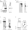
FIGURE 1
FIGURE 1
RITA binds to p53 in cancer cells and in vitro. (A) [14C]-RITA/protein complexes were analyzed in cancer cells treated with 5 μM [14C]-RITA for 12 h to enable sufficient accumulation of RITA. Cell lysates of HCT 116 or HCT 116 TP53 −/− cells were separated in 10% SDS-PAGE under mild denaturing conditions (snap denaturation). The position of [14C]-RITA was visualized by autoradiography. p53 was detected by immunoblotting before and after immunodepletion with DO-1 antibody (right panel). Shown is a representative data of three independent experiments. (B) A small-molecule band shift assay in gradient polyacrylamide gel run under non-denaturing conditions in 4 and 6% TBE gel showed that [14C]-RITA binds to p53 in HCT 116 cells [14C]-RITA and p53 were detected as in Figure A. (C) [14C]-RITA binds to GST-Np53(2–65) fusion protein, and human serum albumin (HSA) but not to fibrinogen as detected by a small-molecule band shift assay in 6% TBE gel using 2:1 M excess of RITA. Dotted line indicates where the gel was cut. (D) Upon standard denaturing conditions [14C]-RITA/p53 and [14C]-RITA/HSA complexes are disrupted.
FIGURE 2
FIGURE 2
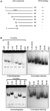
RITA binding site is located between residues 25–38 of the human p53 N-terminus. (A). Scheme depicting the series of deletion mutants generated to map RITA binding site. (B) [14C]-RITA only binds to p53 N terminus deletion mutants containing residues 25–38. Band density in 8% TBE gels was measured using ImageJ software and normalized to GST-tag.
FIGURE 3
FIGURE 3
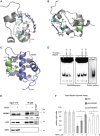
Molecular modelling shows that RITA binds to S33 and S37, induces allosteric shift in Np53 and inhibits p53/MDM2 and p53/MDM4 interactions. (A) Binding of RITA to SPLPS sequence (cyan) of p53 involves interaction with S33 and S37 via terminal hydroxyl groups of RITA, and hydrophobic interactions with P34 and P36. Hydrogen bonds are highlighted in black dotted lines. Orientation of the MDM2-binding helix of p53 (lime) is different upon p53 binding to RITA (blue) (B) and to MDM2 (purple) (pdb: 1YCQ) (C). Side chains of residues (F19, W23 and L26) involved in MDM2 binding are shown in (B,C). Atom type colouring; oxygen (red), nitrogen (blue), and sulphur (yellow). See also Supplementary Movie S1. (D). Proline triad competes for the binding to Np53 with [14C]-RITA in gel shift assay as visualized by autoradiography. CDB3 peptide which binds to p53 core domain does not compete for the binding to Np53. Band density was measured using ImageJ software. (E) In line with the model prediction, RITA-induced p53 conformational change results in the inhibition of p53/MDM2 and p53/MDMX binding in HCT 116 cells as assessed by co-immunoprecipitation. Dotted line indicates the site where the membrane was exposed at different exposure time. (F). RITA rescues the p53 transcription activity from inhibition by MDM2 or MDMX as assessed by yeast-based functional assay. The average light units relative to the transactivation activity of p53 alone and the standard errors of at least five biological repeats are presented. The t-student test was performed for statistical analysis, with p ≤ 0.05.
FIGURE 4
FIGURE 4
Two terminal hydroxyl groups of RITA are crucial for the binding to p53 and the inhibition of the p53/MDM2 interaction. (A) RITA analogue NSC-650973 (compound 4) lacking two hydroxyl groups does not inhibit the growth of HCT 116 cancer cells, unlike LCTA-2081 (compound 2) analogue with substituted O atom in furan ring (for structure refer to Supplementary Table S1), which retained full biological activity. NSC-672170 (compound 3) analogue with one hydroxyl group substituted to ketone retained approximately 60% of RITA biological activity. (B) Compound 4 (40, 80, and 100 µM) does not compete for the binding to Np53 with [14C]-RITA. (C) p53/MDM2 complexes (fluorescent foci) in MCF7 and U2OS cells treated or non-treated with RITA as detected by in situ Proximity Ligation Assay (isPLA). The p53-null H1299 and U2OS cells stained without secondary antibody were used as the assay controls. (D) Quantitative isPLA demonstrated the decrease in the average number of nuclear signals by RITA, but not by its derivative NSC-650973 (cpd 4) (upper panel). The normality was assessed with Shapiro-Wilk’s test. p < 0.05 values were considered statistically significant. RITA, but not compound 4 induced p53 accumulation in HCT116 cells, as detected by western blot (lower panel).
FIGURE 5
FIGURE 5
Serine 33 and serine 37 are crucial for the binding of RITA to the p53 N-terminus. (A) Assessment of [14C]-RITA interaction with Np53 proteins carrying alanine substitutions of S33 or S33/S37 using band shift assay (8% TBE). Np53(S33/S37) binding to [14C]-RITA in vitro is negligible. Bands’ densities were quantified using ImageJ software and normalized to GST-tag. (B) Alignment of murine and human p53 N-termini. Highlighted are the sites for MDM2 interaction and the RITA-binding motif. (C,D) RITA does not bind to mouse Np53 proteins, spanning residues 1–64 and 1–85 as detected by band shift assay in 8% TBE gel and scintillation proximity assay (SPA). Np53(33/37) was used as a negative binding control in SPA assay. (E) Co-immunoprecipitation showed that RITA does not prevent p53/MDM2 interaction in TA3-Sth mouse cancer cells. C—control untreated sample, R—RITA-treated, N—nutlin-treated samples; dotted line represents different exposure time of this part of the membrane. (F). Mouse p53 in Myc- and Ras-transformed mouse embryonic fibroblasts (MEFs) is not induced by RITA (R) in contrast to nutlin (N). (G). RITA induces human p53 in SWAP MEF’s transfected with Ras and c-Myc as detected by western blot.
FIGURE 6
FIGURE 6
Serine 33 and serine 37 are required for RITA-induced rescue of p53 from inhibition by MDM2. (A) Co-expression of MDM2 along with wt or mutant p53(33/37) inhibits p53-dependent luciferase reporter in yeast-based reporter assay. 1 µM RITA (R) does not rescue the reporter driven by mutant p53(33/37). The t-student test was performed for statistical analysis, p < 0.05. N, nutlin. (B) wt p53 and mutant p53(33/37) were overexpressed using lentivirus in RKO TP53 −/− and SW 48 cancer cells. Wt p53 protein but not mutant p53(33/37) is induced by 1 µM RITA (R) as assessed by western blotting. Band density was assessed using ImageJ software and normalized to non-treated controls. N, nutlin. (C) RITA increases the secondary structure content in wt Np53 (left) but not in mutant Np53(33/37) (right) as detected by circular dichroism spectroscopy (CD). (D) nESI mass spectra (left) and drift tube ion mobility-mass spectrometry collision cross section distributions arising from arrival time distributions (right) for the [M + 6H]6+ analyte of wt Np53 in the absence and presence of RITA (top panel, and mutant Np53(33/37) in the absence and presence of RITA (bottom panel). Conformational families are depicted by coloured Gausian curves. wtNp53 undergoes a compaction event resulting in the induction of a novel conformational family shown in red. Mutant Np53(33/37) conformational spread is unaffected by RITA. (E) Protoporphyrin IX, an inhibitor of p53/MDM2 and p53/MDMX interactions, requires serine 33 and serine 37 for binding to Np53 as depicted in a fluorescent pull-down assay. (F) A scheme illustrating allosteric, reversible mechanism of RITA-induced inhibition of p53/MDM2 interaction. Binding of RITA shifts the balance towards p53 conformation with low affinity to MDM2.

References

    1. Belyi V. A., Ak P., Markert E., Wang H., Hu W., Puzio-Kuter A., et al. (2010). The Origins and Evolution of the P53 Family of Genes. Cold Spring Harb. Perspect. Biol. 2, a001198. 10.1101/cshperspect.a001198 - DOI - PMC - PubMed
    1. Bill K. L. J., Garnett J., Meaux I., Ma X., Creighton C. J., Bolshakov S., et al. (2016). SAR405838: A Novel and Potent Inhibitor of the Mdm2:p53 axis for the Treatment of Dedifferentiated Liposarcoma. Clin. Cancer Res. 22, 1150–1160. 10.1158/1078-0432.CCR-15-1522 - DOI - PMC - PubMed
    1. Castell A., Yan Q., Fawkner K., Hydbring P., Zhang F., Verschut V., et al. (2018). A Selective High Affinity MYC-Binding Compound Inhibits MYC:MAX Interaction and MYC-dependent Tumor Cell Proliferation. Sci. Rep. 8, 10064. 10.1038/s41598-018-28107-4 - DOI - PMC - PubMed
    1. Chivian D., Baker D. (2006). Homology Modeling Using Parametric Alignment Ensemble Generation with Consensus and Energy-Based Model Selection. Nucleic Acids Res. 34, e112. 10.1093/nar/gkl480 - DOI - PMC - PubMed
    1. Dawson R., Müller L., Dehner A., Klein C., Kessler H., Buchner J. (2003). The N-Terminal Domain of P53 Is Natively Unfolded. J. Mol. Biol. 332, 1131–1141. 10.1016/j.jmb.2003.08.008 - DOI - PubMed