. 2022 Jun 8:2022:6821170.
doi: 10.1155/2022/6821170. eCollection 2022.

Patchouli Alcohol Inhibits D-Gal Induced Oxidative Stress and Ameliorates the Quality of Aging Cartilage via Activating the Nrf2/HO-1 Pathway in Mice

Affiliations

Patchouli Alcohol Inhibits D-Gal Induced Oxidative Stress and Ameliorates the Quality of Aging Cartilage via Activating the Nrf2/HO-1 Pathway in Mice

Ming Chen et al. Oxid Med Cell Longev. .

Abstract

Chondrocytes play an essential role in maintaining the structure and function of articular cartilage. Oxidative stress occurred in chondrocytes accelerates cell senescence and death, which contributes to the development of osteoarthritis (OA). Patchouli alcohol (PA), a kind of sesquiterpene in Pogostemon cablin, processes multiple bioactivities in treatment of many diseases. However, its effects of antisenescence and antioxidation on chondrocytes in a D-gal-induced aging mice model are still obscure. In this study, we found that PA treatment could ameliorate the degradation of cartilage extracellular matrix (ECM) in a D-gal-induced aging mice model. Further analyses through the immunofluorescent staining and western blot revealed that PA inhibited D-gal-induced chondrocyte senescence via the activation of antioxidative system. Besides, the damage caused by D-gal could not be recovered with PA treatment in Nrf2-silencing chondrocytes. In addition, molecular docking analysis between PA and Keap1 further suggested that the mechanism of PA's antisenescence and antioxidation was attributed to the activation of Nrf2/HO-1 pathway. Therefore, our results demonstrated that PA was a promising candidate for preventing the quality loss of aging cartilage through inhibiting oxidative stress-mediated senescence in chondrocytes.


Conflict of interest statement

The authors declare no conflict of interest.

Figures

Figure 1
Figure 1
Influences of patchouli alcohol (PA) on cartilage quality in D-gal-induced aging mice. (a) Effect of PA on ECM degradation marker gene expression of collagen 2a1 (COL2A1), aggrecan (ACAN), and matrix metalloproteinase 13 (MMP13) in D-gal-caused senescent chondrocytes was analyzed by RT-qPCR. (b) The quantitative real-time PCR (qRT-PCR) results of the mRNA expression of Tp53, Cyclin-Dependent Kinase Inhibitor 1A (Cdkn1a/p21Cip1/Waf1), and Cyclin-Dependent Kinase Inhibitor 2A (Cdkn2a/p16INK4a). (c) Representative alcian blue and safranin O-fast green (SO) staining images in NOR group, D-gal group, and D-gal and PA (20 mg/kg) group. Scale bar, 250 μm; scale bar (enlarged), 100 μm. (d) Immunochemical staining of articular cartilage with COL2A1 and MMP13 in the NOR group, D-gal group, and D-gal and PA (20 mg/kg) group (staining positive areas were circled in red). Scale bar, 100 μm. (e) The diagram was applied to display modified Mankin's score of articular cartilage. (f) Quantitative analysis of COL2A1 and MMP13 immunostaining (positive cells %). Scale bar: 50 μm. Mean ± SEM. P < 0.05 vs. NOR group; #P < 0.05 vs. D-gal group.
Figure 2
Figure 2
Influences of patchouli alcohol (PA) on oxidative stress and Nrf2/HO-1 signaling pathway in D-gal-induced aging mice. (a) The mRNA expression of Kelch-like ECH-associated protein 1 (Keap1) and nuclear factor-erythroid 2-related factor 2 (Nrf2) in knee joint cartilage from aging mice given PA. (b) The mRNA relative abundance of catalase (Cat) and superoxide dismutase 1 (Sod1) in D-gal-mediated senescence mice. (c) The expression of Nrf2 was evaluated using immunofluorescent staining. Scale bar, 100 μm. (d) The activities or contents of serum oxidation activity indicators such as CAT, glutathione (GSH), malondialdehyde (MDA), and SOD were determined using commercial kits. (e) The activities or contents of CAT, GSH, MDA, and SOD in different organs (liver, kidney, spleen, and thymus) were determined by commercial kits. Mean ± SEM. P < 0.05 and ∗∗P < 0.01 vs. NOR group; #P < 0.05 vs. D-gal group.
Figure 3
Figure 3
Influences of patchouli alcohol (PA) on senescence process and extracellular matrix (ECM) homeostasis of chondrocytes induced by D-gal. (a) Effect of PA on cell viability and cytotoxicity was determined by CCK-8 assay. (b) Effect of PA on ECM degradation marker gene expression of collagen 2a1 (Col2a1) and matrix metalloproteinase 13 (Mmp13) in D-gal-caused senescent chondrocytes was analyzed by RT-qPCR. (c) Representative images of alcian blue, safranin O, and SA-β-gal staining in chondrocytes treated with PA (0, 2.5, 5, and 10 μM) and D-gal (5 mg/ml) for 24 h. The scale bar of alcian blue and safranin O staining, 250 μm. The scale bar of SA-β-gal staining, 10 0 μm. (d) Influence of PA on gene expression of Tp53 and Cyclin-Dependent Kinase Inhibitor 1A (Cdkn1a/p21Cip1/Waf1) in D-gal-caused senescent chondrocytes. (e and f) The immunofluorescence detection of MMP13 and TP53 in D-gal-caused senescent chondrocytes was treated with 10 μM PA for 24 h. Scale bar, 50 μm. (g) The expression and quantitative analysis of aggrecan (ACAN), COL2A1, a disintegrin and metalloproteinase with thrombospondin 5 (ADAMTS 5), and MMP13 proteins in chondrocytes disposed with PA (0, 2.5, 5, and 10 μM) and D-gal (5 mg/ml) for 24 h were determined by western blotting. The protein expression level is the gray value ratio of the target protein to β-actin. (h) The expression and quantitative analysis of TP53 and CDKN1A/p21Cip1/Waf1 proteins in D-gal-mediated aging chondrocytes administrated with 10 μM PA for 24 h. The protein expression level is the gray value ratio of the target protein to β-actin. Mean ± SEM. P < 0.05 and ∗∗P < 0.01 vs. control group; #P < 0.05 vs. D-gal group.
Figure 4
Figure 4
Influences of patchouli alcohol (PA) on oxidative stress and expression of Nrf2/HO-1 signaling pathway of chondrocytes mediated by D-gal. (a) Effect of PA on gene expression of catalase (Cat), glutathione synthetase (Gss), and superoxide dismutase 1 (Sod1) in D-gal-caused senescent chondrocytes was analyzed by RT-qPCR. (b) PA's effects upon antioxidant indices (CAT and SOD) generation by chondrocytes administrated with D-gal (5 mg/ml) and PA (0, 2.5, 5, and 10 μM) for 24 h were determined by biochemical kit. (c) Representative fluorescent images of ROS levels in D-gal (5 mg/ml) induced senescent chondrocytes treated with 10 μM PA for 24 h. Scale bar, 50 μm. (d) The RT-qPCR was employed to detect the gene expression of Nrf2/HO-1 pathway, including nuclear factor-erythroid 2-related factor-2 (Nrf2), heme oxygenase-1 (Hmox1), NAD(P)H quinone dehydrogenase 1 (Nqo1), and Kelch-like ECH-associated protein 1 (Keap1), mediated by D-gal in aging chondrocytes. (e) Representative immunofluorescence pictures of Nrf2 were captured in D-gal (5 mg/ml) induced senescent chondrocytes. Nrf2 was stained with Nrf2-specific antibody (red staining), while the nucleus was stained with DAPI (blue staining). Scale bar, 50 μm. (f) The expression and quantitative analysis of Nrf2 protein in the nucleus and HO-1 protein in the cytoplasm were assessed through western blotting. The Nrf2 and HO-1 protein expression levels are the gray value ratio of the target protein to lamin B and β-actin, respectively. Mean ± SEM. P < 0.05 and ∗∗P < 0.01 vs. control group; #P < 0.05 vs. D-gal group.
Figure 5
Figure 5
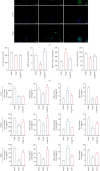
The suppressive effects of patchouli alcohol (PA) on D-gal-induced chondrocyte senescence via upregulating Nrf2/HO-1 signaling pathway. (a–c) Impact of PA on mRNA expression of collagen 2a1 (Col2a1), matrix metalloproteinase 13 (Mmp13), Tp53, Cyclin-Dependent Kinase Inhibitor 1A (Cdkn1a/p21Cip1/Waf1), catalase (Cat), and superoxide dismutase 1 (Sod1) in D-gal-caused senescent chondrocytes was detected by RT-qPCR. (d) Effect of PA on cell viability and cytotoxicity was determined by CCK-8 assay. (e) PA's influence upon antioxidant indices (CAT and SOD) generation by D-gal-induced senescent chondrocytes was determined by biochemical kit. (f) Representative images of alcian blue, safranin O, and SA-β-gal staining in aging chondrocytes. The scale bar of alcian blue and safranin O staining, 250 μm. The scale bar of SA-β-gal staining, 100 μm. (g) Representative fluorescent images of ROS levels in D-gal (5 mg/ml) induced senescent chondrocytes. Scale bar, 50 μm. (h and i) The immunofluorescence detection of MMP13 and TP53 in D-gal-caused senescent chondrocytes. Scale bar, 50 μm. (j) The expression and quantitative analysis of COL2A1, MMP13, nuclear factor-erythroid 2-related factor-2 (Nrf2), heme oxygenase-1 (HO-1), TP53, and p21 proteins were assessed through western blotting. The Nrf2 protein expression level is the gray value ratio of the target protein to lamin B, while other proteins were the grayscale ratio of target protein to β-actin. Mean ± SEM. P < 0.05 and ∗∗P < 0.01 vs. corresponding group.
Figure 6
Figure 6
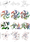
Docking between Nrf2 (1X2J) and three ligands (patchouli alcohol (i), KI696 (ii), and theaflavin (iii)). (a) Molecular structure diagram of patchouli alcohol, KI696, and theaflavin. (b) Global graph of docking results. (c) Local details of 3D molecular docking results. (d) The 2D display of molecular docking details.

Similar articles

Cited by

References

    1. Shin K. R., Kim M. Y., Kim Y. H. Study on the lived experience of aging. Nursing & Health Sciences . 2003;5(4):245–252. doi: 10.1046/j.1442-2018.2003.00161.x. - DOI - PubMed
    1. Argyropoulou A., Aligiannis N., Trougakos I. P., Skaltsounis A. L. Natural compounds with anti-ageing activity. Natural Product Reports . 2013;30(11):1412–1437. doi: 10.1039/c3np70031c. - DOI - PubMed
    1. Campisi J. Aging, cellular senescence, and cancer. Annual Review of Physiology . 2013;75(1):685–705. doi: 10.1146/annurev-physiol-030212-183653. - DOI - PMC - PubMed
    1. Price J. S., Waters J. G., Darrah C., et al. The role of chondrocyte senescence in osteoarthritis. Aging Cell . 2002;1(1):57–65. doi: 10.1046/j.1474-9728.2002.00008.x. - DOI - PubMed
    1. Loeser R. F., Goldring S. R., Scanzello C. R., Goldring M. B. Osteoarthritis: a disease of the joint as an organ. Arthritis and Rheumatism . 2012;64(6):1697–1707. doi: 10.1002/art.34453. - DOI - PMC - PubMed

LinkOut - more resources