. 2022 Jun 10:2022:4107915.
doi: 10.1155/2022/4107915. eCollection 2022.

Inhibition of TRPA1 Ameliorates Periodontitis by Reducing Periodontal Ligament Cell Oxidative Stress and Apoptosis via PERK/eIF2 α/ATF-4/CHOP Signal Pathway

Affiliations

Inhibition of TRPA1 Ameliorates Periodontitis by Reducing Periodontal Ligament Cell Oxidative Stress and Apoptosis via PERK/eIF2 α/ATF-4/CHOP Signal Pathway

Qian Liu et al. Oxid Med Cell Longev. .

Abstract

Objective: In periodontitis, excessive oxidative stress combined with subsequent apoptosis and cell death further exacerbated periodontium destruction. TRPA1, an important transient receptor potential (TRP) cation channel, may participate in the process. This study is aimed at exploring the role and the novel therapeutic function of TRPA1 in periodontitis.

Methods: Periodontal ligament cells or tissues derived from healthy and periodontitis (PDLCs/Ts and P-PDLCs/Ts) were used to analyze the oxidative and apoptotic levels and TRPA1 expression. TRPA1 inhibitor (HC030031) was administrated in inflammation induced by P. gingivalis lipopolysaccharide (P.g.LPS) to investigate the oxidative and apoptotic levels of PDLCs. The morphology of the endoplasmic reticulum (ER) and mitochondria was identified by transmission electron microscope, and the PERK/eIF2α/ATF-4/CHOP signal pathways were detected. Finally, HC030031 was administered to periodontitis mice to evaluate its effect on apoptotic and oxidative levels in the periodontium and the relieving of periodontitis.

Results: The oxidative, apoptotic levels and TRPA1 expression were higher in P-PDLC/Ts from periodontitis patients and in P.g.LPS-induced inflammatory PDLCs. TRPA1 inhibitor significantly decreased the intracellular calcium, oxidative stress, and apoptosis of inflammatory PDLCs and decreased ER stress by downregulating PERK/eIF2α/ATF-4/CHOP pathways. Meanwhile, the overall calcium ion decrease induced by EGTA also exerted similar antiapoptosis and antioxidative stress functions. In vivo, HC030031 significantly reduced oxidative stress and apoptosis in the gingiva and periodontal ligament, and less periodontium destruction was observed.

Conclusion: TRPA1 was highly related to periodontitis, and TRPA1 inhibitor significantly reduced oxidative and apoptotic levels in inflammatory PDLCs via inhibiting ER stress by downregulating PERK/eIF2α/ATF-4/CHOP pathways. It also reduced the oxidative stress and apoptosis in periodontitis mice thus ameliorating the development of periodontitis.


Conflict of interest statement

The authors declare no relevant financial or nonfinancial interests to disclose.

Figures

Figure 1
Figure 1
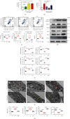
Periodontitis-derived periodontal ligament cells and tissues were at high levels of oxidative stress and apoptosis. (a) The related gene expressions of oxidative stress, apoptosis, and some of the TRP families in healthy and periodontitis-derived periodontal ligament cells (PDLCs and P-PDLCs) (n = 3). (b, c) Western blot and semiquantitative statistical analysis of oxidative stress, apoptosis, and TRPA1 in PDLCs and P-PDLCs. (n = 3). d, Flow cytometry analysis of PDLCs and P-PDLCs (n = 4). (e, f) H&E staining (white star represent immune cell infiltration), immunohistochemistry and immunofluorescence staining, and semiquantitative statistical analysis of periodontitis and healthy derived periodontal ligament tissues (PDLTs and P-PDLTs) (n = 3). Data analysis was performed by using Student's t-test (P < 0.05, ∗∗P < 0.01, and ∗∗∗P < 0.001). The data are presented as the mean ± SEM.
Figure 2
Figure 2
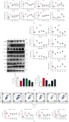
TRPA1 inhibitor HC030031 significantly ameliorated the oxidative stress and apoptosis levels of Pg.LPS-induced PDLCs. (a) P.g.LPS increased intracellular calcium ion level in a dose-dependent manner (n = 3). (b) Lower HC030031 concentration significantly reversed the increase of intracellular calcium ions, and 10 μM HC030031 was used in the subsequent experiment (n = 3). (c) Flow cytometry analysis of the control group (PDLCs only, C), L group (PDLCs treated by LPS, L), and LH group (PDLCs treated by 10 μM HC030031 and LPS, LH) (n = 4). (d, e) Western blot analysis and semiquantitative statistical analysis of oxidative stress, apoptosis, and TRPA1 proteins in in C, L, and LH groups (n = 3). (f) EM images showing endoplasmic reticulum (yellow arrows) and mitochondrial morphology (red arrows) of PDLCs in Ctr, LPS, and LH groups (white stars represent cell nuclei) (n = 4). (g) Quantification of endoplasmic reticulum size, mitochondrial size, mitochondrial number per cell (n = 4), and mitochondrial crista density was analyzed (>100 mitochondria). Data analysis was performed by using one-way ANOVA (P < 0.05, ∗∗P < 0.01, and ∗∗∗P < 0.001). Data are presented as the mean ± SEM.
Figure 3
Figure 3
Immunofluorescence staining of ROS, JC-1, ER-ID, and DHR123 of PDLCs. (a) Immunofluorescence staining of ROS, JC-1, ER-ID, and DHR123 in C, L, LH, LP, LG, and LE groups (n = 4). (b) Semiquantification of immunofluorescence staining (n = 4). Data analysis was performed by using one-way ANOVA (P < 0.05, ∗∗P < 0.01, and ∗∗∗P < 0.001). Error bars represent mean ± SEM.
Figure 4
Figure 4
HC030031 decreased endoplasmic reticulum oxidative stress through the PERK/eIF2α/ATF-4/CHOP pathway. (a) The related gene expressions of PDLCs in the control group (PDLCs only, C), LPS group (PDLCs treated by LPS, L), LH group (PDLCs treated by 10 μM HC030031 and LPS, LH), LP group (PDLCs treated by 10 μM 4-PBA and LPS, LP), LG group (PDLCs treated by 10 μM GSK2656157 and LPS, LG), and LE group (PDLCs treated by 10 μM EGTA and LPS, LE) (n = 4). (b, c) Western blot and semiquantitative statistical analysis of PDLCs in C, L, LH, LP, LG, and LE groups (n = 3). (d) Total SOD and MDAs of PDLCs in C, L, LH, LP, LG, and LE groups (n = 4). (e, f) Flow cytometry and quantitative analysis of PDLCs in C, L, LH, LP, LG, and LE groups (n = 4). Data analysis was performed by using one-way ANOVA and LSD (P < 0.05, ∗∗P < 0.01, and ∗∗∗P < 0.001). Error bars represent mean ± SEM.
Figure 5
Figure 5
TRPA1 inhibitor significantly inhibited periodontal tissue destruction and oxidative stress in periodontitis mice. (a) Representative H&E staining and micro-CT images of mouse maxillary (n = 4): (1) negative control (healthy group intraperitoneally administered with the same solvent but without HC030031, CON); (2) periodontitis group (experimental periodontitis group intraperitoneally administered with the same solvent but without HC030031, Perio); (3) HC1 group (experimental periodontitis group intraperitoneally administered with 10 mg/kg HC030031, HC1); (4) HC3 group (experimental periodontitis group intraperitoneally administered with 30 mg/kg HC030031, HC3). (b) Detailed schematic diagram of the animal experiment process. (c) Quantitative statistical analysis of alveolar bone loss in micro-CT (n = 4). (d) Total SOD and MDAs of mouse serum (n = 4). (e, f) Western blot analysis and semiquantitative statistical analysis of mouse gingiva (n = 4). ABL: alveolar bone loss. Data analysis was performed by using one-way ANOVA (P < 0.05, ∗∗P < 0.01, and ∗∗∗P < 0.001). Error bars represent mean ± SEM.
Figure 6
Figure 6
Oxidative stress and apoptosis levels in gingival tissue and in periodontal ligament tissue were significantly decreased. (a) Immunofluorescence staining of periodontal ligament tissue in mouse maxillary in CON, Perio, HC1, and HC3 groups (n = 4). (b) Semiquantification of immunofluorescence staining (n = 4). Data analysis was performed by using one-way ANOVA (P < 0.05, ∗∗P < 0.01, and ∗∗∗P < 0.001). Error bars represent mean ± SEM.
Figure 7
Figure 7
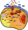
Schematic diagram of the mechanisms of TRPA1 in periodontitis. TRPA1 inhibition ameliorates periodontitis by reducing oxidative stress and apoptosis via PERK/eIF2α/ATF-4/CHOP signal pathway in inflammation.

References

    1. Janakiram C. Dye BA (2020) A public health approach for prevention of periodontal disease. Periodontology . 2000;84(1):202–214. doi: 10.1111/prd.12337. - DOI - PMC - PubMed
    1. Papapanou P. N., Sanz M., Buduneli N., et al. Periodontitis: Consensus report of workgroup 2 of the 2017 World Workshop on the Classification of Periodontal and Peri-Implant Diseases and Conditions. Journal of Clinical Periodontology . 2018;89:S173–S182. doi: 10.1111/jcpe.12946. - DOI - PubMed
    1. Sczepanik F. S. C., Grossi M. L., Casati M., et al. Periodontitis is an inflammatory disease of oxidative stress: We should treat it that way. Periodontology . 2000;84(1):45–68. doi: 10.1111/prd.12342. - DOI - PubMed
    1. Shi L., Ji Y., Zhao S., et al. Crosstalk between reactive oxygen species and Dynamin-related protein 1 in periodontitis. Free radical biology & medicine . 2021;172:19–32. doi: 10.1016/j.freeradbiomed.2021.05.031. - DOI - PubMed
    1. Circu M. L., Aw T. Y. Reactive oxygen species, cellular redox systems, and apoptosis. Free Radical Biology & Medicine . 2010;48(6):749–762. doi: 10.1016/j.freeradbiomed.2009.12.022. - DOI - PMC - PubMed