Review
. 2022 Jun 8:2022:3450207.
doi: 10.1155/2022/3450207. eCollection 2022.

Novel Therapeutic Strategies for Ischemic Stroke: Recent Insights into Autophagy

Affiliations
Review

Novel Therapeutic Strategies for Ischemic Stroke: Recent Insights into Autophagy

Xiaocheng Lu et al. Oxid Med Cell Longev. .

Abstract

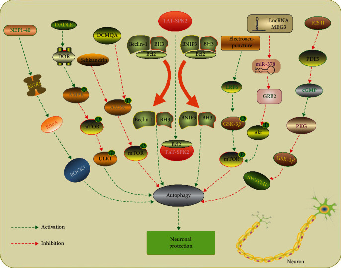
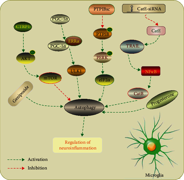
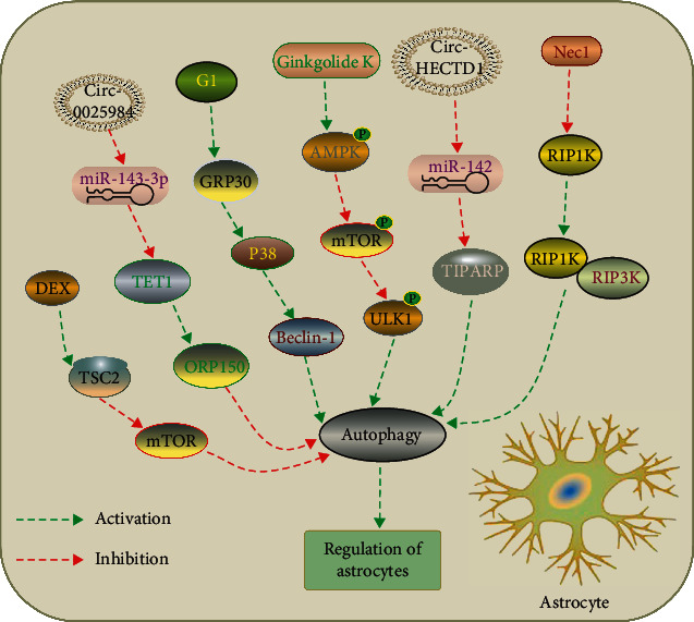
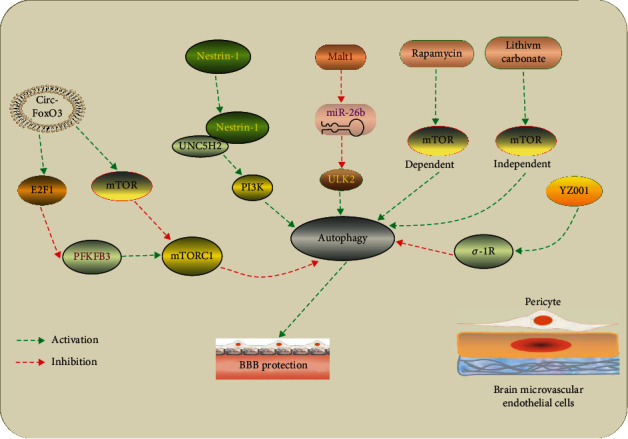
Stroke is one of the leading causes of death and disability worldwide. Autophagy is a conserved cellular catabolic pathway that maintains cellular homeostasis by removal of damaged proteins and organelles, which is critical for the maintenance of energy and function homeostasis of cells. Accumulating evidence demonstrates that autophagy plays important roles in pathophysiological mechanisms under ischemic stroke. Previous investigations show that autophagy serves as a "double-edged sword" in ischemic stroke as it can either promote the survival of neuronal cells or induce cell death in special conditions. Following ischemic stroke, autophagy is activated or inhibited in several cell types in brain, including neurons, astrocytes, and microglia, as well as microvascular endothelial cells, which involves in inflammatory activation, modulation of microglial phenotypes, and blood-brain barrier permeability. However, the exact mechanisms of underlying the role of autophagy in ischemic stroke are not fully understood. This review focuses on the recent advances regarding potential molecular mechanisms of autophagy in different cell types. The focus is also on discussing the "double-edged sword" effect of autophagy in ischemic stroke and its possible underlying mechanisms. In addition, potential therapeutic strategies for ischemic stroke targeting autophagy are also reviewed.


Conflict of interest statement

The authors declare that they have no competing interests.

Figures

Figure 1
Figure 1
Manipulation of autophagy in neurons after ischemic stroke. NEP1-40 treatment inhibits autophagic activation via NGR1/RhoA/ROCK signaling pathway and attenuates secondary neuronal damage [57]. DADLE has been shown to protect ischemic CA1 neurons by activating delta opioid receptor (DOR)-AMPK-autophagy axis [55]. 4 Schizandrin and DCMQA inhibit neuronal apoptosis via suppression of AMPK/mTOR-mediated autophagy [50, 58]. TAT-SPK2 interacts with Bcl-2 via its BH3 domain, thereby dissociating it from Beclin 1, activating autophagy and protecting neurons against ischemic injury [69]. Electroacupuncture pretreatment induces tolerance to cerebral ischemia by inhibiting autophagy through the inhibition of GSK3β [59]. LncRNA MEG3/miR-378/GRB2 axis is involved in neurological functional impairment targeting autophagy in ischemic stroke [60]. Icariside II has been reported to attenuate neuronal injury via inhibiting PKG/GSK-3β/autophagy axis [51].
Figure 2
Figure 2
Manipulation of autophagy in microglia after ischemic stroke. CTRP1 inhibits microglia autophagy and inflammation response by regulating the Akt/mTOR pathway after IS [88]. Microglia-specific PGC-1α activates autophagy through promoting the expression of ULK1 in an ERRα-dependent manner, thereby suppressing neuroinflammation [90]. Inhibition of PTPIB mitigates microglial activation by inhibiting PERK/eIF2α-dependent autophagy after ischemic stroke [87]. A proteolytic relay through the early CatE/TRAIL-dependent proteasomal and late CatB-dependent autophagic pathways for NF-κB activation plays an essential role in the neurotoxic polarization of microglia following ischemic stroke [83]. Geniposide and progesterone inhibits NLRP3 inflammasome activation via suppression autophagy in microglial cells in OGD and ischemic brain injury models [92, 93].
Figure 3
Figure 3
Manipulation of autophagy in astrocytes after ischemic stroke. DEX exerts a neuroprotection against OGD-induced astrocytes injury via activation of astrocytes autophagy by regulating the TSC2/mTOR signaling pathway [108]. Circular RNA 0025984 protects astrocytes from ischemic injury via inhibition of autophagy by targeting the miR-143-3p/TET1/ORP150 pathway [107]. G1 treatment restores autophagy in astrocytes via activation of G protein-coupled receptor 30 (GRP30) and protected neurons after ischemic stroke [109]. Ginkgolide K promotes astrocyte proliferation and migration after oxygen-glucose deprivation via inducing protective autophagy through the AMPK/mTOR/ULK1 signaling pathway [110]. Knockdown of Circular RNA HECTD1 inhibits astrocyte autophagy via MIR142/TIPARP axis, resulting in inhibition of astrocyte activation after cerebral ischemic stroke [106]. Nec-1, a specific inhibitor of RIP1K, decreased RIP1K–RIP3K complex formation and inhibited autophagy, thereby attenuating astrocytic necrotic cell death in the ischemic cortex [105].
Figure 4
Figure 4
Manipulation of autophagy in brain microvascular endothelial cells after ischemic stroke. Circ-FoxO3 inhibits mTORC1 activity mainly by sequestering mTOR and E2F1, thus promoting autophagy to clear cytotoxic aggregates for improving BBB integrity [130]. Netrin-1 ameliorates BBB impairment secondary to ischemic stroke by activating PI3K-mediated autophagy depending on UNC5H2 receptor [129]. LncRNA Malat1 protects brain microvascular endothelial cells (BMECs) against ischemic injury by sponging miR-26b and upregulating ULK2 expression, thereby promoting BMEC autophagy [131]. Rapamycin and lithium carbonate pretreatments improve BBB integrity after ischemic stroke through induction of mTOR-dependent and mTOR-independent autophagy, respectively [127]. YZ001, a new σ-1R agonist, enhances pericyte survival via inhibition of autophagy in ischemic stroke [136].

References

    1. Berkowitz A. L., Mittal M. K., McLane H. C., et al. Worldwide reported use of IV tissue plasminogen activator for acute ischemic stroke. International Journal of Stroke . 2014;9(3):349–355. doi: 10.1111/ijs.12205. - DOI - PubMed
    1. Fischer U., Anca D., Arnold M., et al. Quality of life in stroke survivors after local intra-arterial thrombolysis. Cerebrovasc Dis. . 2008;25(5):438–444. doi: 10.1159/000126917. - DOI - PubMed
    1. California Acute Stroke Pilot Registry. Prioritizing interventions to improve rates of thrombolysis for ischemic stroke. Neurology. . 2005;64(4):654–659. doi: 10.1212/01.WNL.0000151850.39648.51. - DOI - PubMed
    1. De Duve C. The lysosome. Scientific American . 1963;208:64–72. doi: 10.1038/scientificamerican0563-64. - DOI - PubMed
    1. Di Meco A., Curtis M. E., Lauretti E., Pratico D. Autophagy dysfunction in Alzheimer’s disease: mechanistic insights and new therapeutic opportunities. Biological Psychiatry . 2020;87(9):797–807. doi: 10.1016/j.biopsych.2019.05.008. - DOI - PubMed