Masson's Tumor of the Distal Phalanx May Present Like a Felon, Report of a Rare Case
- PMID: 35720211
- PMCID: PMC9201229
- DOI: 10.4103/abr.abr_170_21
Masson's Tumor of the Distal Phalanx May Present Like a Felon, Report of a Rare Case
Abstract
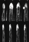
Also known as intravascular papillary endothelial hyperplasia, Masson's tumor is a relatively rare soft-tissue vascular tumor that usually arises in the hand. Felon is an abscess formation in the distal phalanx that usually occurs following a penetrating microtrauma. We present a 30-year-old patient who was referred to our clinic with a palpable mass in the distal phalanx of the index finger after a needle stick injury. At first, the lesion was treated as a felon but finally and after treatment failure, a complete reevaluation revealed the lesion to be a Masson's tumor of the distal phalanx.
Keywords: Distal phalanx; Masson's tumor; felon.
Copyright: © 2022 Advanced Biomedical Research.
Conflict of interest statement
There are no conflicts of interest.
Figures
Similar articles
-
Intravascular papillary endothelial hyperplasia (Masson's tumor) of the finger: a case report and review of the literature.Case Reports Plast Surg Hand Surg. 2025 Jun 5;12(1):2513066. doi: 10.1080/23320885.2025.2513066. eCollection 2025. Case Reports Plast Surg Hand Surg. 2025. PMID: 40487413 Free PMC article.
-
Intravascular Papillary Endothelial Hyperplasia: Case Report of a Recurrent Masson's Tumor of the Finger and Review of Literature.J Hand Microsurg. 2021 Jul;13(3):164-168. doi: 10.1055/s-0039-3401381. Epub 2020 Jan 16. J Hand Microsurg. 2021. PMID: 34602798 Free PMC article.
-
Intraabdominal Intravascular Papillary Endothelial Hyperplasia (Masson's Tumor): A Rare and Novel Cause of Gastrointestinal Bleeding.Case Rep Gastroenterol. 2010 Mar 20;4(1):124-132. doi: 10.1159/000294148. Case Rep Gastroenterol. 2010. PMID: 21103239 Free PMC article.
-
Masson's tumor of the breast: Rare differential for new or recurrent breast cancer-Case report, pathology, and review of the literature.Breast J. 2020 Apr;26(4):752-754. doi: 10.1111/tbj.13608. Epub 2019 Sep 20. Breast J. 2020. PMID: 31538368 Review.
-
Masson's tumor of the hand: a case report and brief literature review.Ann Plast Surg. 2012 Sep;69(3):338-9. doi: 10.1097/SAP.0b013e31822afa63. Ann Plast Surg. 2012. PMID: 21921791 Review.
Cited by
-
Intravascular papillary endothelial hyperplasia (Masson's tumor) of the finger: a case report and review of the literature.Case Reports Plast Surg Hand Surg. 2025 Jun 5;12(1):2513066. doi: 10.1080/23320885.2025.2513066. eCollection 2025. Case Reports Plast Surg Hand Surg. 2025. PMID: 40487413 Free PMC article.
References
-
- Masson P. Hemangioendotheliome vegetant intravasculaire. Bull Soc Anat (Paris) 1923;93:517–23.
-
- Espinosa A, González J, García-Navas F. Intravascular papillary endothelial hyperplasia at foot level: A case report and literature review. J Foot Ankle Surg. 2017;56:72–4. - PubMed
-
- Clifford PD, Temple HT, Jorda M, Marecos E. Intravascular papillary endothelial hyperplasia (Masson's tumor) presenting as a triceps mass. Skeletal Radiol. 2004;33:421–5. - PubMed
-
- Schwartz SA, Taljanovic MS, Harrigal CL, Graham AR, Smyth SH. Intravascular papillary endothelial hyperplasia: Sonographic appearance with histopathologic correlation. J Ultrasound Med. 2008;27:1651–3. - PubMed
-
- Lee SH, Suh JS, Lim BI, Yang WI, Shin KH. Intravascular papillary endothelial hyperplasia of the extremities: MR imaging findings with pathologic correlation. Eur Radiol. 2004;14:822–6. - PubMed
Publication types
LinkOut - more resources
Full Text Sources


