

Case Reports
doi: 10.15386/mpr-1751.
Epub 2022 Jan 31.
Epithelioid angiosarcoma of the masseter muscle: a rare clinicopathological diagnosis
Affiliations
- PMID: 35720232
- PMCID: PMC9177093
- DOI: 10.15386/mpr-1751
Item in Clipboard
Case Reports
Epithelioid angiosarcoma of the masseter muscle: a rare clinicopathological diagnosis
Med Pharm Rep.
2022 Jan.
Abstract
Epithelioid angiosarcoma (EA) is a subtype of angiosarcoma which is a rare tumor of endothelial origin. Here, we report a case of 15-year-old boy who presented with soft tissue mass lesion in the parotid region mimicking as a benign parotid tumor. Cytology was suggestive of inflammatory swelling. Patient underwent superficial parotidectomy along with the wide excision of the swelling. On histopathology, it was diagnosed as EA. To the best of our knowledge this is the first case report of epithelioid angiosarcoma of the masseter.
Keywords: chemotherapy; epithelioid angiosarcoma; masseter muscle; parotid; radiotherapy.
Figures

(a) Markings showing the mass and the modified Blair’s incision in the preauricular region. (b): CT scan showing hypodense lesion occupying the left masseter muscle. (c) Intraoperative picture showing facial nerve and its branches after superficial parotidectomy with small nodular mass in the masseter muscle.
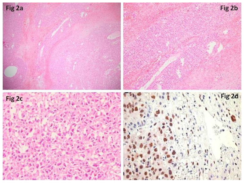
(a,b) Tumor cells are arranged in sheets with interspersed foci of necrosis, congestion, haemorrhage, cystic degeneration and large blood vessels (H&E stain, 10× and 20× respectively). (c) Epithelioid to plump spindle shaped tumor cells having large, moderately pleomorphic round to ovoid nuclei, coarse chromatin, moderate to abundant eosinophilic cytoplasm with few mitotic figures and rich network of small caliber blood vessels are noted (H&E stain, 40×). (d) Immunohistochemistry shows nuclear positivity for FLI-1.
References
-
- Hart J, Mandavilli S. Epithelioid angiosarcoma: a brief diagnostic review and differential diagnosis. Arch Pathol Lab Med. 2011;135:268–272. - PubMed
-
- Weiss SW, Ishak KG, Dail DH, Sweet DE, Enzinger FM. Epithelioid hemangioendothelioma and related lesions. Semin Diagn Pathol. 1986;3:259–287. - PubMed
-
- Ayadi L, Khabir A. Pediatric angiosarcoma of soft tissue: a rare clinicopathologic entity. Arch Pathol Lab Med. 2010;134:481–485. - PubMed
Publication types
LinkOut - more resources
Full Text Sources