. 2022 Jun 1:13:757616.
doi: 10.3389/fimmu.2022.757616. eCollection 2022.

Iguratimod Restrains Circulating Follicular Helper T Cell Function by Inhibiting Glucose Metabolism via Hif1α-HK2 Axis in Rheumatoid Arthritis

Affiliations

Iguratimod Restrains Circulating Follicular Helper T Cell Function by Inhibiting Glucose Metabolism via Hif1α-HK2 Axis in Rheumatoid Arthritis

Ziran Bai et al. Front Immunol. .

Abstract

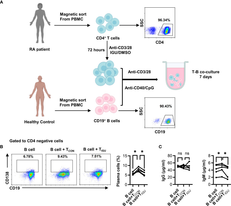
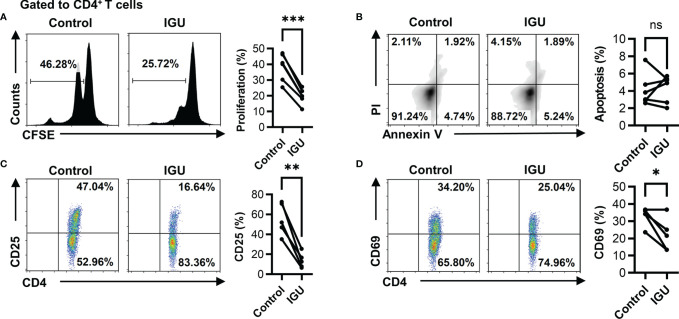
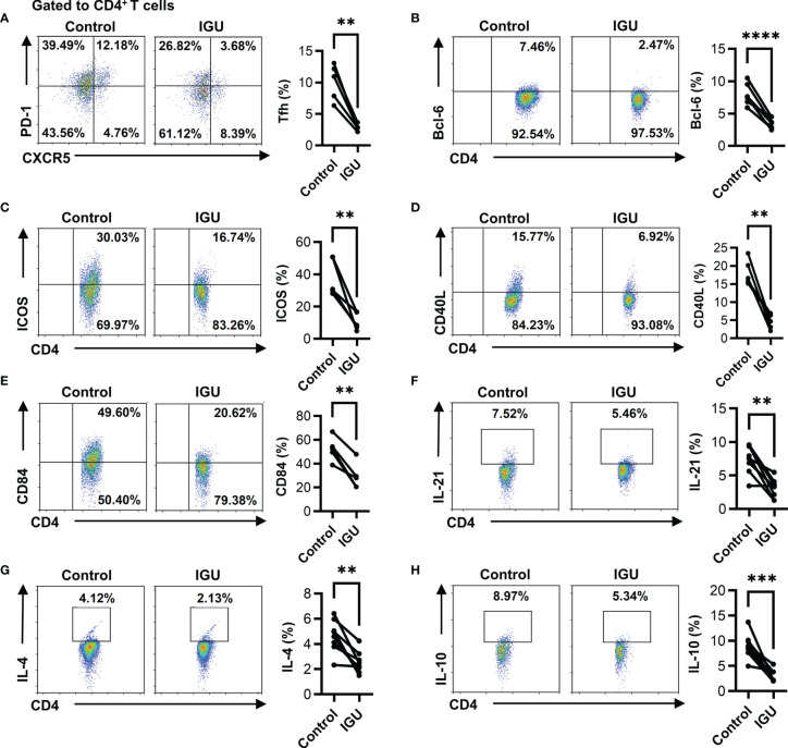
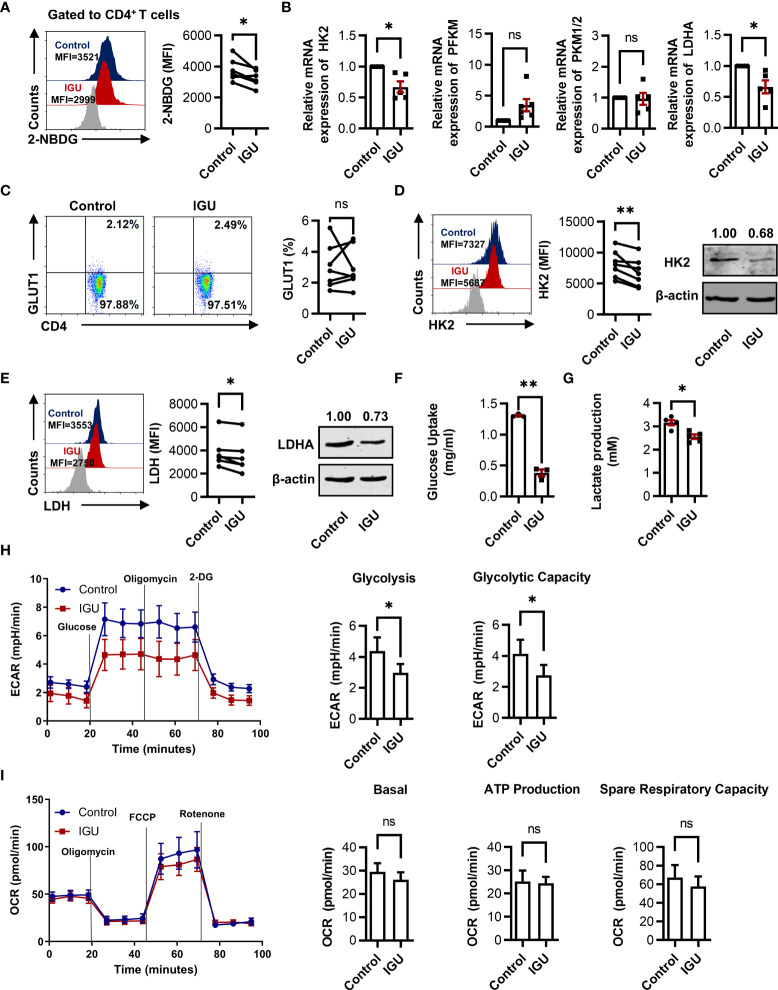
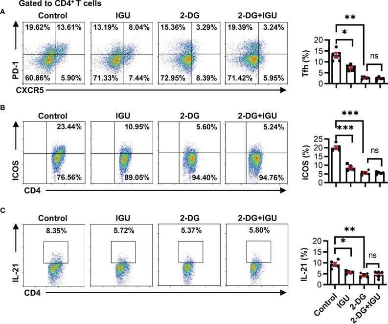
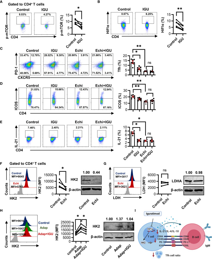
Iguratimod (IGU) is a novel disease modified anti-rheumatic drug, which has been found to act directly on B cells for inhibiting the production of antibodies in rheumatoid arthritis (RA) patients. Follicular helper T (Tfh) cells, a key T cell subsets in supporting B cell differentiation and antibody production, have been shown to play critical roles in RA. However, whether IGU can inhibit RA Tfh cells which further restrains B cell function remains unclear. Here, we aimed to explore the roles of IGU in regulating RA circulating Tfh (cTfh) cell function and investigate the potential mechanism associated with cell glucose metabolism. In our study, we found that IGU could act on RA-CD4+ T cells to reduce T cell-dependent antibody production. IGU decreased the percentage of RA cTfh cells and the expression of Tfh cell-related molecules and cytokines which were involved in B cell functions. Importantly, our data showed that IGU significantly restrained the cTfh cell function by inhibiting glucose metabolism, which relied on Hif1α-HK2 axis. In summary, we clarified a new target and mechanism of IGU by restraining RA cTfh cell function via inhibiting Hif1α-HK2-glucose metabolism axis. Our study demonstrates the potential application of IGU in the treatment of diseases related to abnormal metabolism and function of Tfh cells.

Keywords: Hif1α-HK2 axis; circulating follicular helper T cells; glucose metabolism; iguratimod; rheumatoid arthritis.


Conflict of interest statement

The authors declare that the research was conducted in the absence of any commercial or financial relationships that could be construed as a potential conflict of interest.

Figures

Figure 1
Figure 1
IGU inhibits T cell-dependent antibody production. (A) Purified RA-CD4+ T cells were cultured in the presence or absence of IGU for 72 hours, then co-cultured with CD19+ B cells from healthy donors for 7 days. The purity of CD4+ T cells and CD19+ B cells were shown. (B) The percentage of plasma cells (CD138+) was detected by flow cytometry (n = 5). (C) The production of IgG and IgM in the supernatant was detected by ELISA (n = 5). Symbols represent individual subjects. ns, no significance; *P < 0.05.
Figure 2
Figure 2
IGU inhibits RA-CD4+ T cell proliferation and activation. The PBMCs from RA patients were cultured in the presence of anti-CD3/CD28 antibody (2 μg/ml) in the presence of vehicle (DMSO) or IGU for 5 days. (A) The proliferation of RA-CD4+ T cells was detected by the CFSE labeling method (n = 6). (B) The apoptosis of RA-CD4+ T cells was detected after being cultured for 24 hours (n = 6). Propidium iodide (PI) and Annexin V (AV) staining were determined by flow cytometry. The data of early apoptotic cells (PIAV+) were shown. (C, D) RA-PBMCs were cultured in the presence or absence of IGU for 72 hours. Flow cytometry detected the expression of activation markers (CD25 and CD69) on CD4+ T cells (n = 5). Symbols represent individual subjects. ns, no significance; *P < 0.05; **P < 0.01; ***P < 0.001.
Figure 3
Figure 3
IGU inhibits the cTfh cell function of RA patients. RA PBMCs activated by anti-CD3/CD28 antibody (2 μg/ml) were cultured with IGU or DMSO for 72 hours. (A) The percentage of Tfh (CD4+CXCR5+PD-1+ T) cells was measured by flow cytometry (n = 5). (B) The percentage of CD4+Bcl-6+ T cells was measured by flow cytometry (n = 8). (C–E) The expression of ICOS, CD40L, and CD84 on CD4+ T cells were detected by flow cytometry (n = 5). (F–H) The secreted cytokines of CD4+ T cells, including IL-21, IL-4, and IL-10, were measured by flow cytometry (n = 8). Symbols represent individual subjects. **P < 0.01; ***P < 0.001; ****P < 0.0001.
Figure 4
Figure 4
IGU inhibits glucose metabolism in RA-CD4+ T cells. (A) RA-PBMCs were cultured in the presence of DMSO or IGU for 24 hours, then the glucose uptake was determined by the 2-NBDG method (n = 6). (B) Purified RA-CD4+ T cells were cultured in the presence of IGU or DMSO for 24 hours, and relative mRNA expression levels of HK2, PFKM, PKM1/2, and LDHA were determined by qPCR. Expression levels of each gene were normalized to β-actin expression level and adjusted to the levels in the control group (served as 1). (C-E) GLUT1 (n = 7), HK2 (n = 8), and LDH (n = 6) in RA-CD4+ T cells were measured by flow cytometry and western blotting. (F, G) Purified RA-CD4+ T cells were cultured in the presence of IGU or DMSO for 24 hours. Glucose uptake (n = 3) and lactate production (n = 5) in the culture supernatants were determined by the glucose and lactate assay kit. (H, I) RA-CD4+ T cells were activated by anti-CD3/CD28 for 24 hours, then treated with DMSO or IGU for 1 hour. ECAR (n = 5) and OCR (n = 5) for each group were measured in real-time under basal conditions and after addition of the indicated reagents. The data of glycolysis, glycolytic capacity, basal OCR, ATP production, and spare respiratory capacity were shown. MFI: mean fluorescence intensity. Symbols represent individual subjects. ns, no significance; *P < 0.05; **P<0.01.
Figure 5
Figure 5
IGU restrains the function of cTfh cells by inhibiting HK2. RA-PBMCs were activated by anti-CD3/CD28 antibody (2 μg/ml) and pretreated with HK2 inhibitor 2-DG or not for 4 hours, and then treated with IGU or DMSO for 3 days. (A–C) The frequencies of Tfh cells (A), ICOS+CD4+ T cells (B), and IL-21 producing CD4+ T cells (C) were determined by flow cytometry (n = 5). ns, no significance; *P < 0.05; **P<0.01; ***P < 0.001.
Figure 6
Figure 6
IGU restrains cTfh cell function by inhibiting the Hif1α-HK2 axis. (A, B) RA-PBMCs were activated by anti-CD3/CD28 antibody (2 μg/ml) and treated with or without IGU for 3 days, and the levels of p-mTOR (A) and Hif1α (B) were analyzed by flow cytometry (n = 5). (C–E) RA-PBMCs were pretreated with anti-CD3/CD28 antibody and Hif1α inhibitor Echinimycin (Echi) for 4 hours and then treated with IGU or DMSO for 3 days. The frequencies of Tfh cells (n = 6), ICOS+CD4+ T cells (n = 6), and IL-21 producing CD4+ T cells (n = 4) were determined by flow cytometry. (F, G) RA-PBMCs were treated with Echinomycin (Echi) for 24 hours, HK2 (n = 4) and LDH (n = 4) in RA-CD4+ T cells were measured. (H) RA-PBMCs were pretreated by anti-CD3/CD28 antibody and Adaptaquin (the activator of Hif1α) (5 μM), and then treated with IGU for 72 hours. The expression of HK2 (n = 5) was measured. (I) Schematic depicting the described findings: IGU restrains RA cTfh cell function by inhibiting the Hif1α-HK2 axis. MFI, mean fluorescence intensity. ns, no significance; *P < 0.05; **P<0.01.

Similar articles

Cited by

References

    1. Scott DL, Wolfe F, Huizinga TW. Rheumatoid Arthritis. Lancet (2010) 376(9746):1094–108. doi: 10.1016/S0140-6736(10)60826-4 - DOI - PubMed
    1. Iwata S, Nakayamada S, Fukuyo S, Kubo S, Yunoue N, Wang SP, et al. . Activation of Syk in Peripheral Blood B Cells in Patients With Rheumatoid Arthritis: A Potential Target for Abatacept Therapy. Arthritis Rheumatol (2015) 67(1):63–73. doi: 10.1002/art.38895 - DOI - PubMed
    1. Ji LS, Sun XH, Zhang X, Zhou ZH, Yu Z, Zhu XJ, et al. . Mechanism of Follicular Helper T Cell Differentiation Regulated by Transcription Factors. J Immunol Res (2020) 2020:1826587. doi: 10.1155/2020/1826587 - DOI - PMC - PubMed
    1. Jiang H, Gao H, Wang Q, Wang M, Wu B. Molecular Mechanisms and Clinical Application of Iguratimod: A Review. BioMed Pharmacother (2020) 122:109704. doi: 10.1016/j.biopha.2019.109704 - DOI - PubMed
    1. Xie S, Li S, Tian J, Li F. Iguratimod as a New Drug for Rheumatoid Arthritis: Current Landscape. Front Pharmacol (2020) 11:73. doi: 10.3389/fphar.2020.00073 - DOI - PMC - PubMed

Publication types