. 2022 May 27;24(1):230.
doi: 10.3892/ol.2022.13351. eCollection 2022 Jul.

Nogo-A/NgR signaling regulates stemness in cancer stem-like cells derived from U87MG glioblastoma cells

Affiliations

Nogo-A/NgR signaling regulates stemness in cancer stem-like cells derived from U87MG glioblastoma cells

Chengjin Ai et al. Oncol Lett. .

Abstract

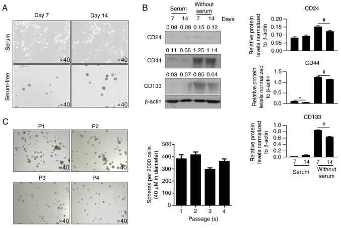
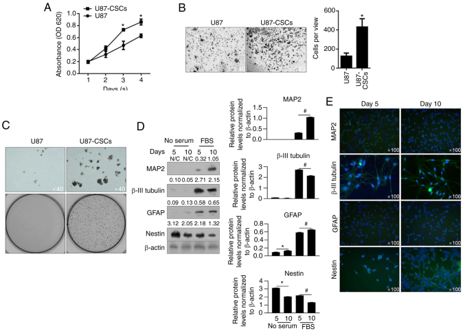
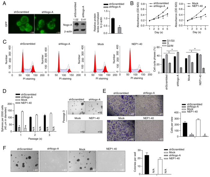
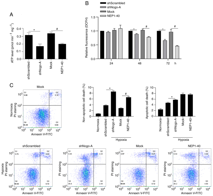
Neurite outgrowth inhibitor A (Nogo-A), a member of the reticulon 4 family, is an axon regeneration inhibitor that is negatively associated with the malignancy of oligodendroglial tumors. It has been suggested that the Nogo-A/Nogo Receptor (NgR) pathway plays a promoting effect in regulating cancer stem-like cells (CSCs) derived from glioblastoma, indicating that Nogo-A could exert different roles in CSCs than those in parental cancer cells. In the present study, CSCs were generated from the human Uppsala 87 malignant glioma (U87MG) cell line. These U87MG-CSCs were characterized by the upregulation of CD44 and CD133, which are two markers of stemness. The expression levels of Nogo-A and the differentiation of U87MG-CSCs were investigated. In addition, the proliferation, invasion and colony formation U87MG-CSCs were examined. Using culture in serum-containing medium, U87MG-CSCs were differentiated into neuron-like cells specifically expressing MAP2, β-III-tubulin and nestin. Nogo-A was upregulated in U87MG-CSCs compared with parental cells. Knockdown of Nogo-A and inhibition of the Nogo-A/NgR signaling pathway in U87MG-CSCs markedly decreased cell viability, cell cycle entry, invasion and tumor formation, indicating that Nogo-A could regulate U87MG-CSC function. Moreover, Nogo-A was involved in intracellular ATP synthesis and scavenging of accumulated reactive oxygen species. Nogo-A/NgR pathway exerted protective effects against hypoxia-induced non-apoptotic and apoptotic cell death. These results suggest that Nogo-A plays an important role in regulating U87MG-CSCs via the Nogo-A/NgR signaling pathway. Nogo-A may also different roles in U87MG-CSCs compared with their parental cells.

Keywords: Nogo-A; apoptosis; cancer stem-like cells; glioblastoma; malignancy.


Conflict of interest statement

The authors declare that they have no competing interests.

Figures

Figure 1.
Figure 1.
Generation and characterization of U87MG-CSCs. (A) After 7 and 14 days of culture with or without serum, cells were imaged. (B) Western blot analysis of CD24, CD44 and CD133 levels. *P<0.05 vs. 7 days with serum. #P<0.05 vs. 7 days without serum. (C) Serial replating assays were performed to detect the stemness of U87MG-CSCs. CSCs, cancer stem cells.
Figure 2.
Figure 2.
Proliferation, invasion and colony formation of U87 CSCs and its parental cells. (A) By performing CCK-8 assays, the proliferation of U87MG-CSCs from day 1 to 4 was measured. *P<0.05 vs. U87MG parental cells. (B) Transwell assays were performed to detect invasiveness of U87MG-CSCs. *P<0.05 vs. U87MG parental cells. (C) Tumor formation in soft agar was employed to detect the tumor formation in U87MG-CSCs compared to their parental cells. Upper lane, Magnification, ×40; lower lane, imaged by digital camera. (D) To analyze differentiation, CSCs were cultured in FBS-supplemented medium for 5 and 10 days, then analyzed by western blotting. *P<0.05 vs. 5 days without serum; #P<0.05 vs. 5 days with serum. (E) Immunofluorescence staining of the neuronal markers, MAP2, β-III tubulin, GFAP and nestin. CCK-8, Cell Counting Kit-8; CSCs, cancer stem cells; MAP2, microtubule-associated protein 2; GFAP, glial fibrillary acidic protein; CSCs, cancer stem cells.
Figure 3.
Figure 3.
Nogo-A is upregulated in CSCs compared with parental cells. (A) Nogo-A expression was validated using GEPIA server (http://gepia.cancer-pku.cn/) in several type of cancer. (B) Western blotting was performed to detect Nogo-A in U87MG and U87MG-CSCs from passage 1 to 4. *P<0.05, vs. U87MG parental cells. (C) Western blotting was performed to detect Nogo-A after differentiation in serum-containing medium for 5 and 10 days. *P<0.05 vs. serum-free group; #P<0.05 vs. 10% FBS group. Nogo-A, neurite outgrowth inhibitor A; GEPIA, Gene Expression Profiling Interactive Analysis; Nogo-A, neurite outgrowth inhibitor A; CSCs, cancer stem cells; BRCA, breast invasive carcinoma; BLCA, bladder urothelial carcinoma.
Figure 4.
Figure 4.
Nogo-A regulates malignant behaviors and stemness in U87-CSCs via Nogo-A/NgR signaling pathway. (A) GFP signal in vector was imaged to confirm the successful transfection of shScrambled or shNogo-A (left panel). Nogo-A was detected to confirm the knockdown efficacy of Nogo-A protein level (right panel). *P<0.05, vs. shScrambled. (B) After Nogo-A knockdown or inhibition of Nogo-A/NgR signaling pathway, cell viability from day 1 to 5 was measured by CCK-8. (C) Cell cycle distribution was measured by PI staining followed by flow cytometry. *P<0.05 vs. shScrambled; #P<0.05 vs. mock. (D) Serial replating assays were measured from passage 1 to 3. *P<0.05 vs. shScrambled; #P<0.05 vs. mock. (E) Transwell assays were performed. *P<0.05 vs. shScrambled. #P<0.05 vs. mock. (F) Tumor formation was performed in soft agar. GFP, green fluorescent protein; Nogo-A, neurite outgrowth inhibitor A; CCK-8, Cell Counting Kit-8; NgR, Nogo Receptor. CSCs, cancer stem cells; PI, propidium iodide.
Figure 5.
Figure 5.
Nogo-A is related to mitochondrial function and protects U87MG-CSCs from hypoxia-induced cell death. (A) Synthesis of ATP under normoxia. *P<0.05 vs. shScrambled; #P<0.05 vs. mock. (B) ROS accumulation at 24, 48 and 72 h. *P<0.05 vs. shScrambled; #P<0.05 vs. mock. (C) Non-apoptotic cell death and apoptotic cell death levels were measured under hypoxic conditions by performing Annexin V-FITC and PI double staining. Quadrant 1 corresponds to non-apoptotic cell death; quadrants 2 and 3 correspond to apoptotic cell death. *P<0.05 vs. shScrambled/hypoxia group; #P<0.05 vs. mock/hypoxia group. Nogo-A, neurite outgrowth inhibitor A; PI, propidium iodide; DCFH, dichlorodihydrofluorescein.

Similar articles

Cited by

References

    1. Mitre AO, Florian AI, Buruiana A, Boer A, Moldovan I, Soritau O, Florian SI, Susman S. Ferroptosis involvement in glioblastoma treatment. Medicina (Kaunas) 2022;58:319. doi: 10.3390/medicina58020319. - DOI - PMC - PubMed
    1. Wang Y, Jiang T. Understanding high grade glioma: Molecular mechanism, therapy and comprehensive management. Cancer Lett. 2013;331:139–146. doi: 10.1016/j.canlet.2012.12.024. - DOI - PubMed
    1. Ramaswamy V, Taylor MD. The amazing and deadly glioma race. Cancer Cell. 2015;28:275–277. doi: 10.1016/j.ccell.2015.08.010. - DOI - PubMed
    1. Alcantara Llaguno S, Parada LF. Cancer stem cells in gliomas: Evolving concepts and therapeutic implications. Curr Opin Neurol. 2021;34:868–874. doi: 10.1097/WCO.0000000000000994. - DOI - PMC - PubMed
    1. Lathia JD, Mack SC, Mulkearns-Hubert EE, Valentim CL, Rich JN. Cancer stem cells in glioblastoma. Genes Dev. 2015;29:1203–1217. doi: 10.1101/gad.261982.115. - DOI - PMC - PubMed