Review
. 2022 Jun 1:13:910228.
doi: 10.3389/fpls.2022.910228. eCollection 2022.

Molecular Mechanisms of Plant Trichome Development

Affiliations
Review

Molecular Mechanisms of Plant Trichome Development

Guoliang Han et al. Front Plant Sci. .

Abstract

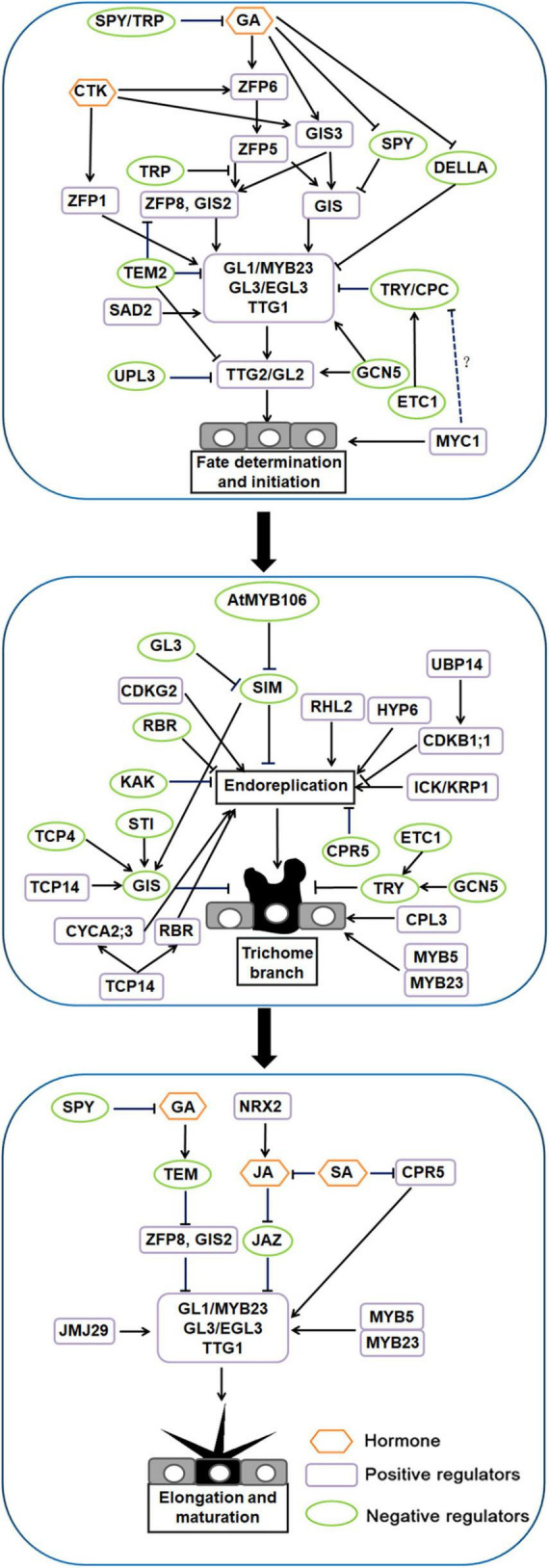
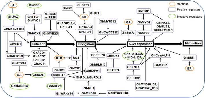
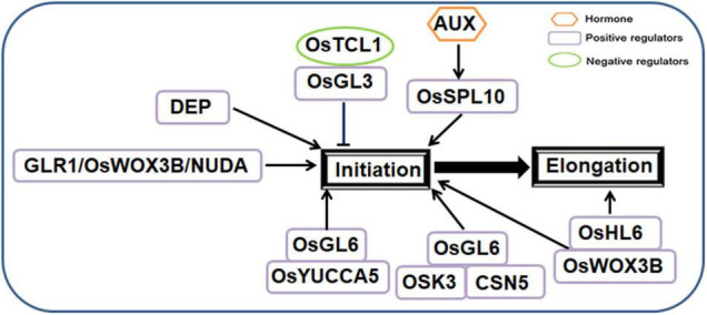
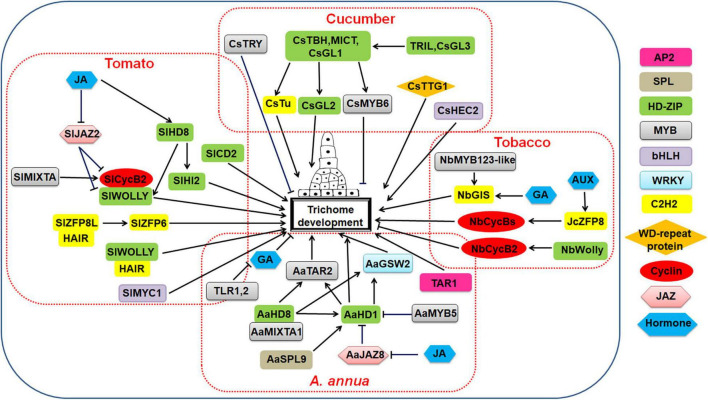
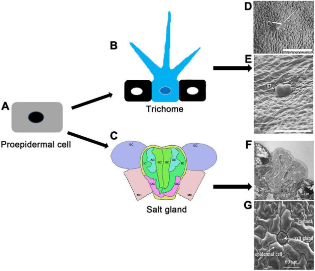
Plant trichomes, protrusions formed from specialized aboveground epidermal cells, provide protection against various biotic and abiotic stresses. Trichomes can be unicellular, bicellular or multicellular, with multiple branches or no branches at all. Unicellular trichomes are generally not secretory, whereas multicellular trichomes include both secretory and non-secretory hairs. The secretory trichomes release secondary metabolites such as artemisinin, which is valuable as an antimalarial agent. Cotton trichomes, also known as cotton fibers, are an important natural product for the textile industry. In recent years, much progress has been made in unraveling the molecular mechanisms of trichome formation in Arabidopsis thaliana, Gossypium hirsutum, Oryza sativa, Cucumis sativus, Solanum lycopersicum, Nicotiana tabacum, and Artemisia annua. Here, we review current knowledge of the molecular mechanisms underlying fate determination and initiation, elongation, and maturation of unicellular, bicellular and multicellular trichomes in several representative plants. We emphasize the regulatory roles of plant hormones, transcription factors, the cell cycle and epigenetic modifications in different stages of trichome development. Finally, we identify the obstacles and key points for future research on plant trichome development, and speculated the development relationship between the salt glands of halophytes and the trichomes of non-halophytes, which provides a reference for future studying the development of plant epidermal cells.

Keywords: development; epidermal cell; molecular mechanism; plant; trichome.


Conflict of interest statement

The authors declare that the research was conducted in the absence of any commercial or financial relationships that could be construed as a potential conflict of interest.

Figures

FIGURE 1
FIGURE 1
Regulatory network model for trichome development in Arabidopsis.
FIGURE 2
FIGURE 2
Regulatory network model for trichome (fiber) development in cotton.
FIGURE 3
FIGURE 3
Regulatory network model for trichome development in rice.
FIGURE 4
FIGURE 4
Regulatory network model for multicellular trichome development in cucumber, tomato, tobacco, and Artemisia annua.
FIGURE 5
FIGURE 5
Possible evolutionary patterns in trichome and salt gland development (Breuer et al., 2009; Yuan et al., 2015b,2016a; Chalvin et al., 2020). (A) Schematic diagram of plant proepidermal cells; (B) schematic diagram of plant trichome; (C) schematic diagram of multicellular salt gland in Limonium bicolor. SC, secretory cell; AC, accessory cell; IC, inner cup cell; OC, outer cup cell; MC, mesophyll cell; EC, epidermal cell; (D) scanning electron microscope observation of Arabidopsis trichomes, 10-day-old wild-type [Columbia (Col)] on the first true leaf; (E) the multicellular glandular trichomes of A. annua leaf (adverse) were observed by scanning electron microscope. GT, glandular trichome. Scale bar = 100 μm. (F) Longitudinal section of salt glands in Limonium bicolor. Transmission electron microscope (TEM) image of salt glands of Limonium bicolor leaves prepared by high-pressure freezing (HPF) followed by freeze substitution (FS) and then embedded, sectioned, and stained. Scale bar = 25 μm; (G) morphologies of salt glands in Limonium bicolor leaves using environmental scanning electron microscopy (ESEM). Scale bar = 60 μm.

Similar articles

Cited by

References

    1. Aashima K., Paper J. M., Boehler A. P., Bradley A. M., Neumann T. R., Kathrin S. (2014). HD-Zip Proteins GL2 and HDG11 Have Redundant Functions in Arabidopsis Trichomes, and GL2 Activates a Positive Feedback Loop via MYB23. Plant Cell 26 2184–2200. 10.1105/tpc.113.120360 - DOI - PMC - PubMed
    1. Akhtar M. Q., Qamar N., Yadav P., Kulkarni P., Kumar A., Shasany A. K. (2017). Comparative glandular trichome transcriptome-based gene characterization reveals reasons for differential (–)-menthol biosynthesis in Mentha species. Physiol. Plant. 160 128–141. 10.1111/ppl.12550 - DOI - PubMed
    1. Allen E., Xie Z., Gustafson A. M., Carrington J. C. (2005). Microrna-Directed phasing during trans -acting siRNA Biogenesis in Plants. Cell 121 207–221. 10.1016/j.cell.2005.04.004 - DOI - PubMed
    1. Amme S., Rutten T., Melzer M., Sonsmann G., Vissers J. P. C., Schlesier B., et al. (2005). A proteome approach defines protective functions of tobacco leaf trichomes. Proteomics 5 2508–2518. 10.1002/pmic.200401274 - DOI - PubMed
    1. An L. J., Zhou Z. J., Su S., Yan A., Gan Y. B. (2012). GLABROUS INFLORESCENCE STEMS (GIS) is required for trichome branching through gibberellic acid signaling in Arabidopsis. Plant. Cell Physiol. 53 457–469. 10.1093/pcp/pcr192 - DOI - PubMed

LinkOut - more resources