Review
. 2022 Jun 2:16:861480.
doi: 10.3389/fnins.2022.861480. eCollection 2022.

Automatic Detection of High-Frequency Oscillations With Neuromorphic Spiking Neural Networks

Affiliations
Review

Automatic Detection of High-Frequency Oscillations With Neuromorphic Spiking Neural Networks

Karla Burelo et al. Front Neurosci. .

Abstract

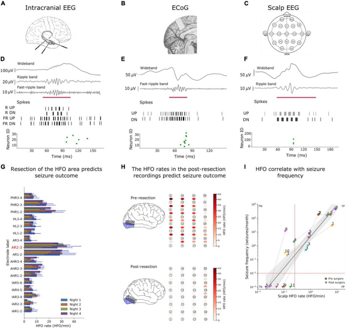
Interictal high-frequency oscillations (HFO) detected in electroencephalography recordings have been proposed as biomarkers of epileptogenesis, seizure propensity, disease severity, and treatment response. Automatic HFO detectors typically analyze the data offline using complex time-consuming algorithms, which limits their clinical application. Neuromorphic circuits offer the possibility of building compact and low-power processing systems that can analyze data on-line and in real time. In this review, we describe a fully automated detection pipeline for HFO that uses, for the first time, spiking neural networks and neuromorphic technology. We demonstrated that our HFO detection pipeline can be applied to recordings from different modalities (intracranial electroencephalography, electrocorticography, and scalp electroencephalography) and validated its operation in a custom-designed neuromorphic processor. Our HFO detection approach resulted in high accuracy and specificity in the prediction of seizure outcome in patients implanted with intracranial electroencephalography and electrocorticography, and in the prediction of epilepsy severity in patients recorded with scalp electroencephalography. Our research provides a further step toward the real-time detection of HFO using compact and low-power neuromorphic devices. The real-time detection of HFO in the operation room may improve the seizure outcome of epilepsy surgery, while the use of our neuromorphic processor for non-invasive therapy monitoring might allow for more effective medication strategies to achieve seizure control. Therefore, this work has the potential to improve the quality of life in patients with epilepsy by improving epilepsy diagnostics and treatment.

Keywords: electroencephalography; epilepsy; epileptogenic tissue; neuromorphic system; spiking neural networks; system-on-a-chip.


Conflict of interest statement

The authors declare that the research was conducted in the absence of any commercial or financial relationships that could be construed as a potential conflict of interest.

Figures

FIGURE 1
FIGURE 1
Example of an HFO. (A) HFO in iEEG signal as recorded. (B) HFO in iEEG filtered in the Ripple band (80–250 Hz) and Fast-ripple band (250–500 Hz). The gray line indicates the presence of an HFO. (C) The time-frequency transform of the filtered signal shows the HFO as an isolated peak. (D) Raster plot measured from the artificial neurons of our SNN. Multiple neurons spiked (blue dots) as they recognized the presence of the HFO [Modified from Sharifshazileh et al. (2021)].
FIGURE 2
FIGURE 2
The SNN to detect HFO in iEEG, ECoG and the scalp EEG. The input to our SNNs are the UP and DN spike trains generated by converting the signals into spikes. The core SNN architecture (green box, used for iEEG) for HFO detection consists of input neurons (gray) receiving the input UP-DN spikes from the filtered signal in HFO band (ripple band and FR band for iEEG, FR band for ECoG, and ripple band for scalp EEG). These inputs project to a second layer of neurons (green) with different synaptic parameters. The core SNN can also interact with the in-band artifact rejection SNN (purple box, added for ECoG). For this interaction, the inputs of the core SNN are projected to the dis-inhibitory neuron (purple) using excitatory synapses. This neuron projects inhibitory synapses to a global-inhibitory neuron (orange), which is continuously inhibiting the second layer neurons. The role of the interneuron and the inhibitory neuron is to avoid the false detection of sharp transients. The artifact rejection SNN (yellow box, added for scalp EEG) consists of input neurons (gray) receiving the input UP-DN spikes from filtering the signal above 500 Hz. These inputs project to a second layer of neurons (yellow) with different synaptic parameters. Panels modified from Burelo et al. (2021, and Sharifshazileh et al. (2021).
FIGURE 3
FIGURE 3
Examples of HFO detection in iEEG, ECoG and scalp EEG. (A–C) We detected HFO in three recording modalities. (A) We analyzed iEEG recordings from a patient with temporal lobe epilepsy (TLE) who was implanted with depth electrodes, (B) the ECoG from a patient whose surgery was guided using ECoG electrodes and (c) a scalp EEG from a child with drug-resistant focal lesional epilepsy. [(D–F) upper panels] The wideband signal was filtered in HFO bands [(D–F) upper panels], The filtered signals were converted into spikes [(D–F) middle panel] and sent as input to the SNNs. The green dots of the raster plots [(D–F) bottom panels] show the activity of the second layer neurons indicating the presence of an HFO in the signal, and the purple mark shows the time window that our detector marked as HFO due to this spiking activity. (G) We used the HFO rates found in iEEG to define the HFO area (AR2-3), which was compare to the resection area to predict the seizure outcome of the patient. (H) Our SNN found a high HFO rate (HFO rate > 1 HFO/min) in the pre-resection ECoG from a patient. The SNN found a HFO rate of > 1HFO/min in a patient who suffers from recurrent seizures. Hence, our SNN predicted seizure recurrence in the individual patient. (I) The scalp HFO rate correlated with seizure frequency of our pediatric patient and mirrored the surgical treatment response. Panels modified from Burelo et al. (2021, , Sharifshazileh et al. (2021).
FIGURE 4
FIGURE 4
Overview of the hardware implementation of our HFO detection system. (A) Abstract schematic of the pre-processing and configuration pipeline. Signals from the electrodes pass through amplification, filtering and analog delta modulation (ADM) delta modulation to reach the spiking neural network (SNN). The chip can be configured, send and receive data through an FPGA daughterboard and via a personal computer; note that this is not required to be online during the operation of the chip. (B) Micrograph of the eight channels of analog headstage implemented on the top-left corner of the chip (the image is rotated 90° clockwise) and they are located right next to one of the four neural cores.

References

    1. Andrade-Valenca L. P., Dubeau F., Mari F., Zelmann R., Gotman J. (2011). Interictal scalp fast oscillations as a marker of the seizure onset zone. Neurology 77 524–531. 10.1212/WNL.0b013e318228bee2 - DOI - PMC - PubMed
    1. Barba C., Cross J. H., Braun K., Cossu M., Klotz K. A., De Masi S., et al. (2020). Trends in pediatric epilepsy surgery in Europe between 2008 and 2015: country-, center-, and age-specific variation. Epilepsia 61 216–227. 10.1111/epi.16414 - DOI - PubMed
    1. Bartolozzi C., Indiveri G. (2007). Synaptic dynamics in analog VLSI. Neural Comput. 19 2581–2603. 10.1162/neco.2007.19.10.2581 - DOI - PubMed
    1. Bartolozzi C., Indiveri G., Donati E. (2022). Embodied neuromorphic intelligence. Nat. Commun. 13. 10.1038/s41467-022-28487-2 - DOI - PMC - PubMed
    1. Boran E., Ramantani G., Krayenbuhl N., Schreiber M., Konig K., Fedele T., et al. (2019b). High-density ECoG improves the detection of high frequency oscillations that predict seizure outcome. Clin. Neurophysiol. 130 1882–1888. 10.1016/j.clinph.2019.07.008 - DOI - PubMed

LinkOut - more resources