Diagnostic Performance of Diffusion Kurtosis Imaging for Benign and Malignant Breast Lesions: A Systematic Review and Meta-Analysis
- PMID: 35721236
- PMCID: PMC9203235
- DOI: 10.1155/2022/2042736
Diagnostic Performance of Diffusion Kurtosis Imaging for Benign and Malignant Breast Lesions: A Systematic Review and Meta-Analysis
Retraction in
-
Retracted: Diagnostic Performance of Diffusion Kurtosis Imaging for Benign and Malignant Breast Lesions: A Systematic Review and Meta-Analysis.Appl Bionics Biomech. 2023 Nov 29;2023:9810302. doi: 10.1155/2023/9810302. eCollection 2023. Appl Bionics Biomech. 2023. PMID: 38075141 Free PMC article.
Abstract
Purpose: Magnetic resonance imaging (MRI) has a high sensitivity for differentiating between malignant and non-malignant breast lesions but is sometimes limited due to its low specificity. Here, we performed a meta-analysis to evaluate the diagnostic performance of mean kurtosis (MK) and mean diffusivity (MD) values in magnetic resonance diffusion kurtosis imaging (DKI) for benign and malignant breast lesions.
Methods: Original articles on relevant topics, published from 2010 to 2019, in PubMed, EMBASE, and WanFang databases were systematically reviewed. According to the purpose of the study and the characteristics of DKI reported, the diagnostic performances of MK and MD were evaluated, and meta-regression was conducted to explore the source of heterogeneity.
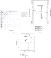
Results: Fourteen studies involving 1,099 (451 benign and 648 malignant) lesions were analyzed. The pooled sensitivity, pooled specificity, positive likelihood ratio, and negative likelihood ratio for MD were 0.84 (95% confidence interval (CI), 0.81-0.87), 0.83 (95% CI, 0.79-0.86), 4.44 (95% CI, 3.54-5.57), and 0.18 (95% CI, 0.13-0.26), while those for MK were 0.89 (95% CI, 0.86-0.91), 0.86 (95% CI, 0.82-0.89), 5.72 (95% CI, 4.26-7.69), and 0.13 (95% CI, 0.09-0.19), respectively. The overall area under the curve (AUC) was 0.91 for MD and 0.95 for MK.
Conclusions: Analysis of the data from 14 studies showed that MK had a higher pooled sensitivity, pooled specificity, and diagnostic performance for differentiating between breast lesions, compared with MD.
Copyright © 2022 Hongyu Gu et al.
Conflict of interest statement
The authors declare that they have no competing interests.
Figures


References
Publication types
LinkOut - more resources
Full Text Sources

