

Biomechanical Properties of Paraspinal Muscles Influence Spinal Loading-A Musculoskeletal Simulation Study
- PMID: 35721854
- PMCID: PMC9201424
- DOI: 10.3389/fbioe.2022.852201
Biomechanical Properties of Paraspinal Muscles Influence Spinal Loading-A Musculoskeletal Simulation Study
Abstract
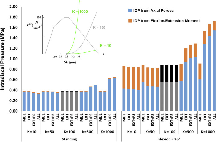
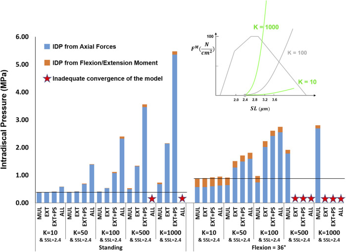
Paraspinal muscles are vital to the functioning of the spine. Changes in muscle physiological cross-sectional area significantly affect spinal loading, but the importance of other muscle biomechanical properties remains unclear. This study explored the changes in spinal loading due to variation in five muscle biomechanical properties: passive stiffness, slack sarcomere length (SSL), in situ sarcomere length, specific tension, and pennation angle. An enhanced version of a musculoskeletal simulation model of the thoracolumbar spine with 210 muscle fascicles was used for this study and its predictions were validated for several tasks and multiple postures. Ranges of physiologically realistic values were selected for all five muscle parameters and their influence on L4-L5 intradiscal pressure (IDP) was investigated in standing and 36° flexion. We observed large changes in IDP due to changes in passive stiffness, SSL, in situ sarcomere length, and specific tension, often with interesting interplays between the parameters. For example, for upright standing, a change in stiffness value from one tenth to 10 times the baseline value increased the IDP only by 91% for the baseline model but by 945% when SSL was 0.4 μm shorter. Shorter SSL values and higher stiffnesses led to the largest increases in IDP. More changes were evident in flexion, as sarcomere lengths were longer in that posture and thus the passive curve is more influential. Our results highlight the importance of the muscle force-length curve and the parameters associated with it and motivate further experimental studies on in vivo measurement of those properties.
Keywords: biomechanics; intradiscal pressure; lumbar spine; muscle; musculoskeletal model; passive stiffness; sarcomere.
Copyright © 2022 Malakoutian, Sanchez, Brown, Street, Fels and Oxland.
Conflict of interest statement
The authors declare that the research was conducted in the absence of any commercial or financial relationships that could be construed as a potential conflict of interest.
Figures


Similar articles
-
The effect of vertebral level on biomechanical properties of the lumbar paraspinal muscles in a rat model.J Mech Behav Biomed Mater. 2021 Jun;118:104446. doi: 10.1016/j.jmbbm.2021.104446. Epub 2021 Mar 15. J Mech Behav Biomed Mater. 2021. PMID: 33780860
-
Dysfunctional paraspinal muscles in adult spinal deformity patients lead to increased spinal loading.Eur Spine J. 2022 Sep;31(9):2383-2398. doi: 10.1007/s00586-022-07292-x. Epub 2022 Jul 16. Eur Spine J. 2022. PMID: 35842491 Free PMC article.
-
Subject-specific biomechanics of trunk: musculoskeletal scaling, internal loads and intradiscal pressure estimation.Biomech Model Mechanobiol. 2016 Dec;15(6):1699-1712. doi: 10.1007/s10237-016-0792-3. Epub 2016 May 12. Biomech Model Mechanobiol. 2016. PMID: 27169402
-
Loads distributed in vivo among vertebrae, muscles, spinal ligaments, and intervertebral discs in a passively flexed lumbar spine.Biomech Model Mechanobiol. 2020 Dec;19(6):2015-2047. doi: 10.1007/s10237-020-01322-7. Epub 2020 Apr 20. Biomech Model Mechanobiol. 2020. PMID: 32314072
-
The effect of posture on lumbar muscle morphometry from upright MRI.Eur Spine J. 2020 Sep;29(9):2306-2318. doi: 10.1007/s00586-020-06409-4. Epub 2020 Apr 25. Eur Spine J. 2020. PMID: 32335742
Cited by
-
Spinal cord injury modeling: from modeling to evaluation using rats as examples.Front Neurol. 2025 Jun 16;16:1573779. doi: 10.3389/fneur.2025.1573779. eCollection 2025. Front Neurol. 2025. PMID: 40589986 Free PMC article. Review.
-
Lumbar muscle adaptations to external perturbations are modulated by trunk posture.Eur J Appl Physiol. 2023 Oct;123(10):2191-2202. doi: 10.1007/s00421-023-05223-2. Epub 2023 May 29. Eur J Appl Physiol. 2023. PMID: 37247004
-
Advances in Musculoskeletal Modeling of the Thoraco-Lumbar Spine: A Comprehensive Systematic Review.Ann Biomed Eng. 2025 Sep 5. doi: 10.1007/s10439-025-03818-8. Online ahead of print. Ann Biomed Eng. 2025. PMID: 40913215 Review.
-
A Muscle-Driven Spine Model for Predictive Simulations in the Design of Spinal Implants and Lumbar Orthoses.Bioengineering (Basel). 2025 Mar 6;12(3):263. doi: 10.3390/bioengineering12030263. Bioengineering (Basel). 2025. PMID: 40150727 Free PMC article.
-
The effects of spinal flexion exposure on lumbar muscle shear modulus and posture.Eur J Appl Physiol. 2025 Jan;125(1):175-182. doi: 10.1007/s00421-024-05586-0. Epub 2024 Aug 19. Eur J Appl Physiol. 2025. PMID: 39158592
References
LinkOut - more resources
Full Text Sources