. 2022 Sep:7:100015.
doi: 10.1016/j.immuno.2022.100015. Epub 2022 Jun 11.

Epitope identification of SARS-CoV-2 structural proteins using in silico approaches to obtain a conserved rational immunogenic peptide

Affiliations

Epitope identification of SARS-CoV-2 structural proteins using in silico approaches to obtain a conserved rational immunogenic peptide

Leonardo Pereira de Araújo et al. Immunoinformatics (Amst). 2022 Sep.

Abstract

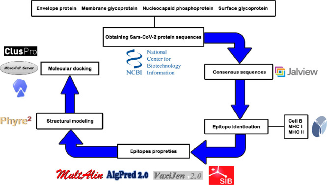
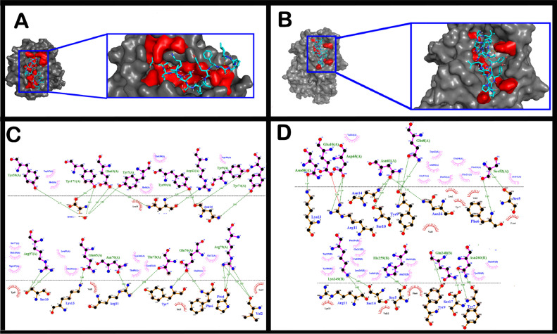
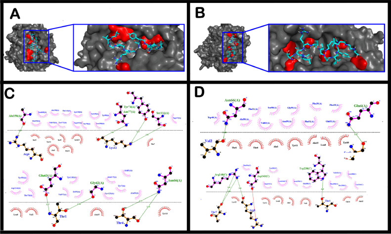
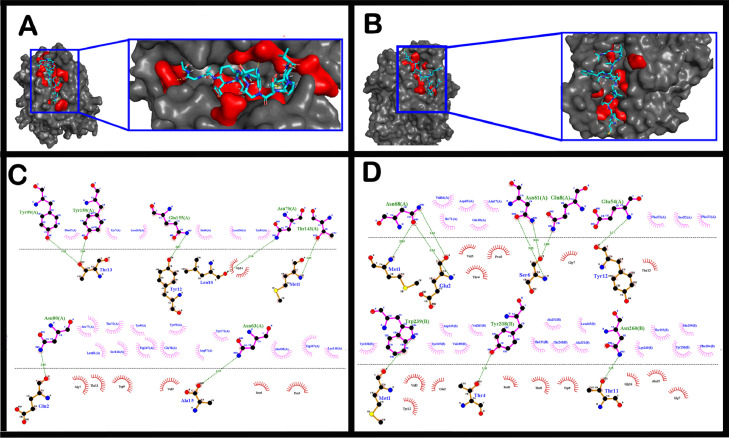
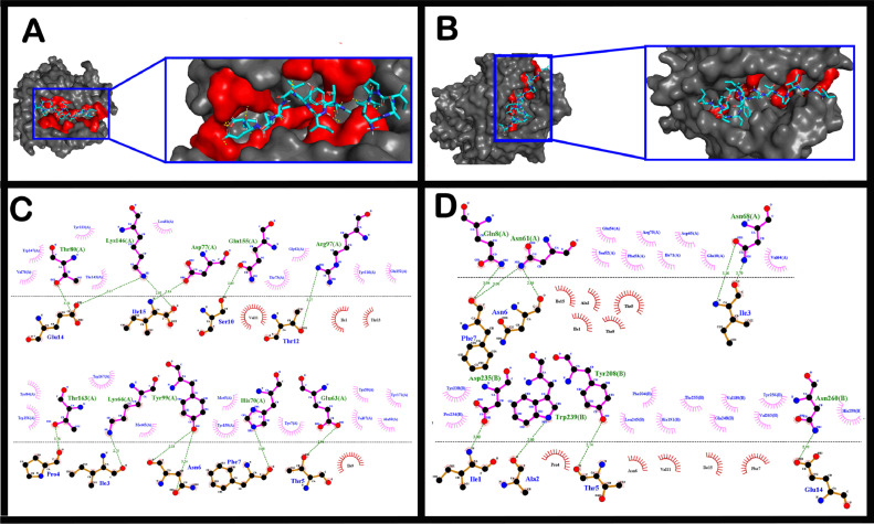
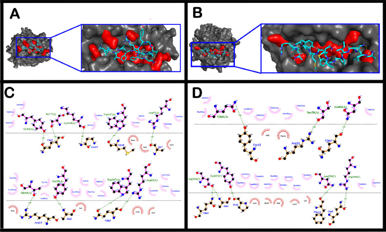
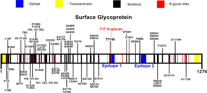
The short time between the first cases of COVID-19 and the declaration of a pandemic initiated the search for ways to stop the spread of SARS-CoV-2. There are great expectations regarding the development of effective vaccines that protect against all variants, and in the search for it, we hypothesized the obtention of a predicted rational immunogenic peptide from structural components of SARS-CoV-2 might help the vaccine research direction. In the search for a candidate of an immunogenic peptide of the SARS-CoV-2 envelope (E), membrane (M), nucleocapsid (N), or spike (S) proteins, we access the predicted sequences of each protein after the genome sequenced worldwide. We obtained the consensus amino acid sequences of about 14,441 sequences of each protein of each continent and the worldwide consensus sequence. For epitope identification and characterization from each consensus structural protein related to MHC-I or MHC-II interaction and B-cell receptor recognition, we used the IEDB reaching 68 epitopes to E, 174 to M, 245 to N, and 833 to S proteins. To select an epitope with the highest probability of binding to the MHC or BCR, all epitopes of each consensus sequence were aligned. The curation indicated 1, 4, 8, and 21 selected epitopes for E, M, N, and S proteins, respectively. Those epitopes were tested in silico for antigenicity obtaining 16 antigenic epitopes. Physicochemical properties and allergenicity evaluation of the obtained epitopes were done. Ranking the results, we obtained one epitope of each protein except for the S protein that presented two epitopes after the selection. To check the 3D position of each selected epitope in the protein structure, we used molecular homology modeling. Afterward, each selected epitope was evaluated by molecular docking to reference MHC-I or MHC-II allelic protein sequences. Taken together, the results obtained in this study showed a rational search for a putative immunogenic peptide of SARS-CoV-2 structural proteins that can improve vaccine development using in silico approaches. The epitopes selected represent the most conserved sequence of new coronavirus and may be used in a variety of vaccine development strategies since they are also presented in the described variants of SARS-CoV-2.

Keywords: COVID-19; New coronavirus; Reverse vaccinology; SARS-CoV-2.


Conflict of interest statement

The authors declare that they have no known competing financial interests or personal relationships that could have appeared to influence the work reported in this paper.

Figures

Image, graphical abstract
Graphical abstract
Fig. 1
Fig. 1
Position identification of selected epitopes in consensus 3D structural proteins from SARS-CoV-2. Molecular modeling by homology for the envelope protein (A), membrane glycoprotein (B), nucleocapsid phosphoprotein (C), and surface glycoprotein (D). The molecular modeling of the proteins was based on homology using AlphaFold v2.0 scrip. The selected epitopes are highlighted in red, and the transmembrane helices are indicated in the models in green.
Fig. 2
Fig. 2
Interaction between the selected epitope from the consensus sequence of the SARS-CoV-2 envelope protein and MHC-I and MHC-II alleles. The best interaction evaluated by molecular docking between the envelope protein epitope and the MHC-I allele HLA-B*1501(A and C) and MHC-II allele HLA-DRB1*04:01 (B and D). The points of contact are demonstrated in A and B in red, and the points of interaction between the epitope and MHC molecules are 2D represented in C and D, respectively.
Fig. 3
Fig. 3
Interaction between the selected epitope from the consensus sequence of the SARS-CoV-2 membrane glycoprotein and MHC-I and MHC-II alleles. The best interaction evaluated by molecular docking between the membrane glycoprotein epitope and the MHC-I allele HLA-B*57:01 (A and C), and MHC-II allele HLA-DRB1*15:01 (B and D). The points of contact are demonstrated in A and B in red, and the points of interaction between the epitope and MHC molecules are 2D represented in C and D, respectively.
Fig. 4
Fig. 4
Interaction between the selected epitope from the consensus sequence of the SARS-CoV-2 nucleocapsid phosphoprotein and MHC-I and MHC-II alleles. The best interaction evaluated by molecular docking between the nucleocapsid phosphoprotein epitope and the MHC-I allele HLA-B*35:01 (A and C), and MHC-II allele HLA-DRB1*04:01 (B and D). The points of contact are demonstrated in A and B in red, and the points of interaction between the epitope and MHC molecules are 2D represented in C and D, respectively.
Fig. 5
Fig. 5
Interaction between the selected epitope 1 from the SARS-CoV-2 surface glycoprotein consensus sequence and MHC-I and MHC-II alleles. The best interaction evaluated by molecular docking between the surface glycoprotein epitope 1 and the MHC-I allele HLA-A*02:03 (A and C) and MHC-II allele HLA-DRB1*04:01 (B and D). The points of contact are demonstrated in A and B in red in red, and the points of interaction between the epitope and MHC molecules are 2D represented in C and D, respectively.
Fig. 6
Fig. 6
Interaction between the selected epitope 2 from the SARS-CoV-2 surface glycoprotein consensus sequence and MHC-I and MHC-II alleles. The best interaction evaluated by molecular docking between the surface glycoprotein epitope 2 and the MHC-I allele HLA-B*35:01 (A and C), and MHC-II allele HLA-DRB1*15:01 (B and D). The points of contact are demonstrated in A and B in red, and the points of interaction between the epitope and MHC molecules are 2D represented in C and D, respectively.
Fig. 7
Fig. 7
SARS-CoV-2 surface glycoprotein variants. The described variants of SARS-CoV-2 based on amino acid changes in the surface glycoprotein indicate a variety of points across the protein indicated by a red mark. The selected epitopes are highlighted in blue. N-glycan sites are highlighted in green. The selected epitope 1 lies within a described variant T716I and a putative n-glycan site, while epitope 2 is not included within any yet-described variant.

Similar articles

Cited by

References

    1. WORLD HEALTH ORGANIZATION. WHO announces COVID-19 outbreak a pandemic. March 12, 2020. https://www.euro.who.int/en/health-topics/health-emergencies/coronavirus... (accessed 19 july 2021). Reference to a dataset:.
    1. WU Di, et al. The SARS-CoV-2 outbreak: what we know. Int J Infect Dis. 2020;94:44–48. doi: 10.1016/j.ijid.2020.03.004. - DOI - PMC - PubMed
    1. PETERSEN Eskild, et al. Comparing SARS-CoV-2 with SARS-CoV and influenza pandemics. Lancet Infect Dis. 2020 doi: 10.1016/S1473-3099(20)30484-9. - DOI - PMC - PubMed
    1. Yao XH, Luo T, et al. A cohort autopsy study defines COVID-19 systemic pathogenesis. Cell Res. 2021 Jun 16 doi: 10.1038/s41422-021-00523-8. - DOI - PMC - PubMed
    1. UL QAMAR, Tahir Muhammad, et al. Reverse vaccinology assisted designing of multiepitope-based subunit vaccine against SARS-CoV-2. Infect Dis Poverty. 2020;9(1):1–14. doi: 10.1186/s40249-020-00752-w. a. - DOI - PMC - PubMed