. 2022 Jun 1:13:915069.
doi: 10.3389/fmicb.2022.915069. eCollection 2022.

Human Virus Genomes Are Enriched in Conserved Adenine/Thymine/Uracil Multiple Tracts That Pause Polymerase Progression

Affiliations

Human Virus Genomes Are Enriched in Conserved Adenine/Thymine/Uracil Multiple Tracts That Pause Polymerase Progression

Emanuela Ruggiero et al. Front Microbiol. .

Abstract

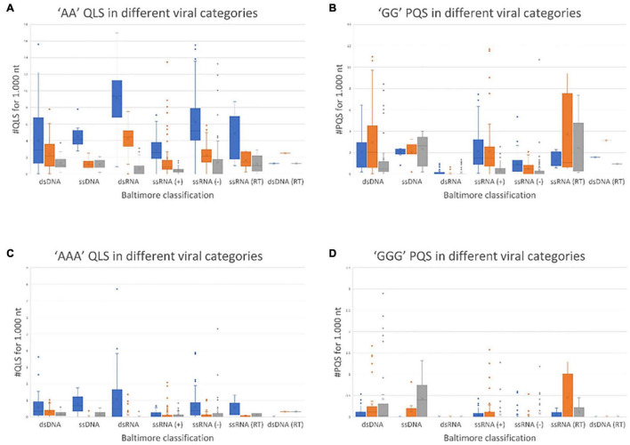
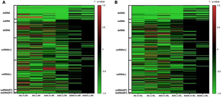
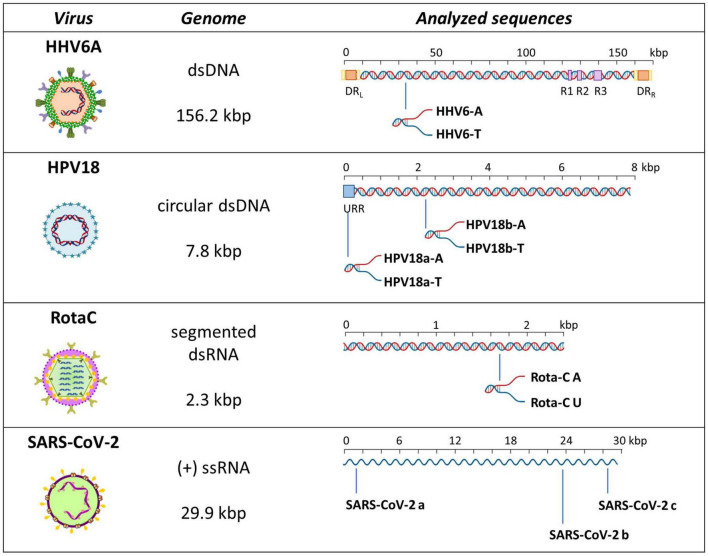
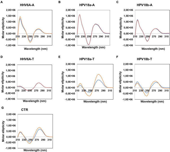
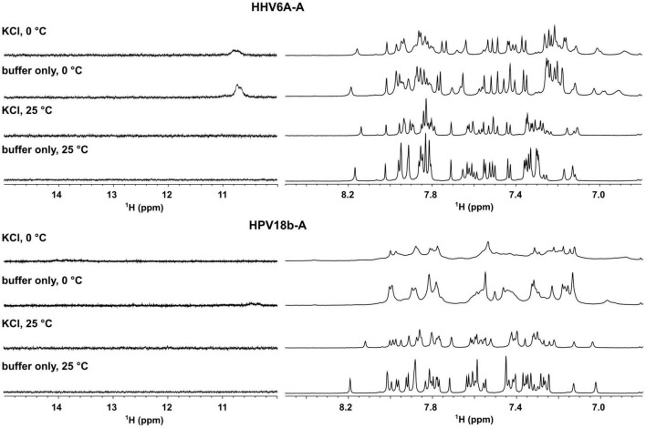
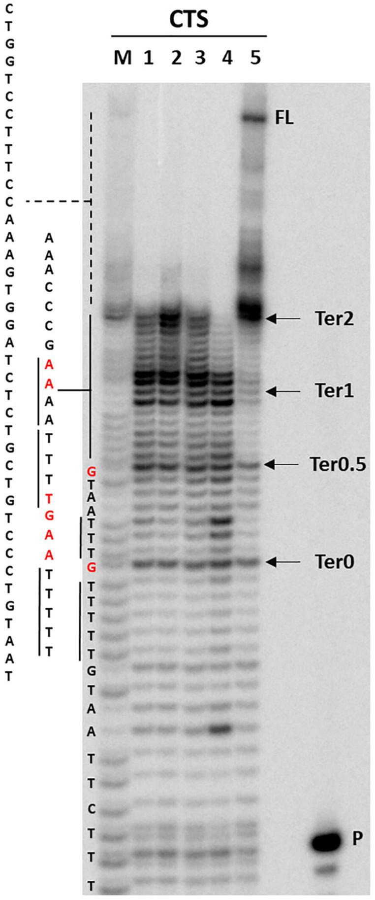
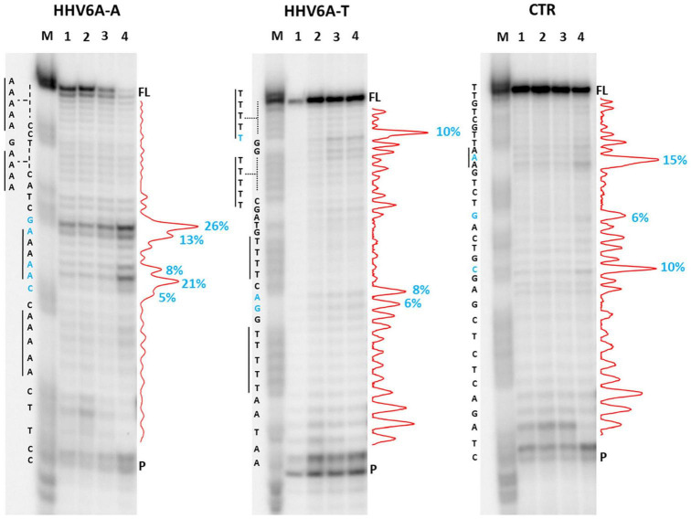
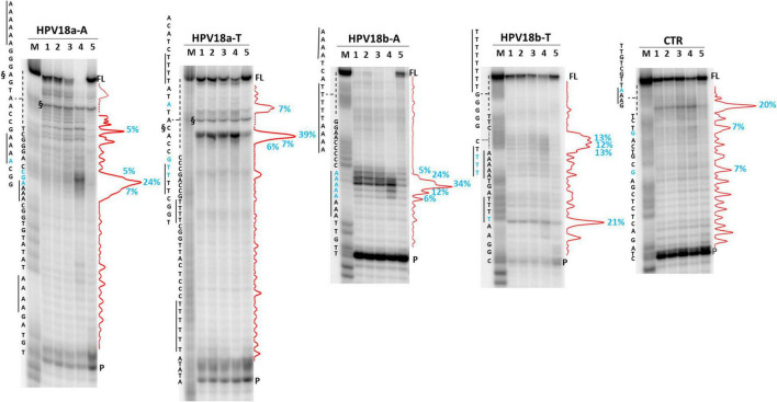
The DNA secondary structures that deviate from the classic Watson and Crick base pairing are increasingly being reported to form transiently in the cell and regulate specific cellular mechanisms. Human viruses are cell parasites that have evolved mechanisms shared with the host cell to support their own replication and spreading. Contrary to human host cells, viruses display a diverse array of nucleic acid types, which include DNA or RNA in single-stranded or double-stranded conformations. This heterogeneity improves the possible occurrence of non-canonical nucleic acid structures. We have previously shown that human virus genomes are enriched in G-rich sequences that fold in four-stranded nucleic acid secondary structures, the G-quadruplexes.Here, by extensive bioinformatics analysis on all available genomes, we showed that human viruses are enriched in highly conserved multiple A (and T or U) tracts, with such an array that they could in principle form quadruplex structures. By circular dichroism, NMR, and Taq polymerase stop assays, we proved that, while A/T/U-quadruplexes do not form, these tracts still display biological significance, as they invariably trigger polymerase pausing within two bases from the A/T/U tract. "A" bases display the strongest effect. Most of the identified A-tracts are in the coding strand, both at the DNA and RNA levels, suggesting their possible relevance during viral translation. This study expands on the presence and mechanism of nucleic acid secondary structures in human viruses and provides a new direction for antiviral research.

Keywords: adenines; conservation; non-canonical structures; polymerase progression; viruses.


Conflict of interest statement

The authors declare that the research was conducted in the absence of any commercial or financial relationships that could be construed as a potential conflict of interest.

Figures

FIGURE 1
FIGURE 1
Normalized abundance of A-QLSs (A,C) and G-PQSs (B,D) in the genome of all human viruses, grouped by Baltimore class. Each panel refers to the specified type of island (AA and AAA for A-QLSs, GG and GGG for G-PQSs); boxplots are delimited by the first and third quartile and the inside horizontal line is the median value of the QLS distribution. Whiskers delimit all the points that fall above/below the third/first quartile ± 1.5 times of the interquartile range (IQR). Bar colors indicate the strand where the predicted patterns are found with respect to coding sequences and refer to concordant (same strand of a CDS, blue), discordant (opposite strand of a CDS, orange), and non-coding (no CDS overlaps that pattern, gray), respectively.
FIGURE 2
FIGURE 2
Comparison of A-QLS abundance in real viral genomes vs. simulated genomes reshuffled at (A) single nucleotide and (B) island levels. Green and red indicate enrichment in real and simulated sequences, respectively.
FIGURE 3
FIGURE 3
Schematic representation of analyzed sequences. For each sequence, the relative virus, its genome features, and a schematic genome organization are reported. Adapted from Ruggiero and Richter (2018).
FIGURE 4
FIGURE 4
Circular dichroism analysis of HHV6A-A (A), HPV18b-A (B), and HPV-18b-A (C), and respective complementary T-rich sequences (D–F). Samples were prepared in the absence (blue line) or presence (red line) of 100 mM KCl. The CTR sequence (G) was used as a control for unstructured DNA. Analysis was performed at 20°C.
FIGURE 5
FIGURE 5
Aromatic (right) and imino (left) region of 1D proton NMR spectra of HHV6A-A, and HPV18b-A oligonucleotides in lithium cacodylate buffer with and without 100 mM KCl. Spectra were recorded at 25 and 0°C.
FIGURE 6
FIGURE 6
Taq polymerase stop assay on HIV-1 CTS sequence. Analysis was performed in the absence (lane 5) or presence of 100 mM different salt types, such a KCl (1), NaCl (2), LiCl (3), and NaF (4). P indicates the unreacted labeled primer; FL indicates the full-length elongation product. Arrows indicate the previously reported termination sites (Lavigne et al., 1997). Bases relative to stop sites are reported in red.
FIGURE 7
FIGURE 7
Taq polymerase stop assay on HHV6A sequences. Analysis was performed in the presence of 100 mM of different salt types, such as KCl (1), NaCl (2), LiCl (3), and NaF (4). M indicates a sequence marker lane, obtained by the Maxam and Gilbert sequencing protocol; P indicates the unreacted labeled primer; FL indicates the full-length elongation product. Red densitograms show stop bands quantification. Percentages of peaks (≥5%) are reported in blue, along with the corresponding nucleotide. Elongation was performed at 37°C.
FIGURE 8
FIGURE 8
Taq polymerase stop assay on HPV-18 sequences. Analysis was performed in the absence (lane 5) or presence of 100 mM different salt types, such as KCl (1), NaCl (2), LiCl (3), and NaF (4). M indicates a sequence marker lane, obtained by the Maxam and Gilbert sequencing protocol; P indicates the unreacted labeled primer; FL indicates the full-length elongation product; § indicates primer dimer band and was excluded from quantitation (dotted line). Red densitograms show stop bands quantification. Percentages of peaks (≥5%) are reported in blue, along with the corresponding nucleotide. Elongation was performed at 37°C.
FIGURE 9
FIGURE 9
RT stop assay on RotaC (A) and SARS-CoV-2 (B) sequences. Analysis was performed in the absence (lane 5) or presence of 100 mM different salt types, such as KCl (1), NaCl (2), LiCl (3), and NaF (4). M indicates a sequence marker lane, obtained by the Maxam and Gilbert sequencing protocol; P indicates the unreacted labeled primer; FL indicates the full-length elongation product; § indicates primer dimer band and was excluded from quantitation (dotted line). Red densitograms show stop bands quantification. Percentages of peaks (≥5%) are reported in blue, along with the corresponding nucleotide. Elongation was performed at 44°C.

Similar articles

References

    1. Ablasser A., Bauernfeind F., Hartmann G., Latz E., Fitzgerald K. A., Hornung V. (2009). RIG-I-dependent sensing of poly(dA:dT) through the induction of an RNA polymerase III-transcribed RNA intermediate. Nat. Immunol. 10 1065–1072. 10.1038/ni.1779 - DOI - PMC - PubMed
    1. Alexeev D. G., Lipanov A. A., Skuratovskii I. Y. (1987). Poly(dA)-poly(dT) is a B-type double helix with a distinctively narrow minor groove. Nature 325 821–823. 10.1038/325821a0 - DOI - PubMed
    1. Behe M. J. (1995). An overabundance of long oligopurine tracts occurs in the genome of simple and complex eukaryotes. Nucleic Acids Res. 23 689–695. 10.1093/nar/23.4.689 - DOI - PMC - PubMed
    1. Berselli M., Lavezzo E., Toppo S. (2020). QPARSE: searching for long-looped or multimeric G-quadruplexes potentially distinctive and druggable. Bioinformatics 36 393–399. 10.1093/bioinformatics/btz569 - DOI - PubMed
    1. Bhattacharyya D., Arachchilage G. M., Basu S. (2016). Metal cations in G-quadruplex folding and stability. Front. Chem. 4:38. 10.3389/fchem.2016.00038 - DOI - PMC - PubMed

LinkOut - more resources