. 2022 May;10(10):540.
doi: 10.21037/atm-22-2068.

Electroacupuncture of the Baihui and Shenting acupoints for vascular dementia in rats through the miR-81/IL-16/PSD-95 pathway

Affiliations

Electroacupuncture of the Baihui and Shenting acupoints for vascular dementia in rats through the miR-81/IL-16/PSD-95 pathway

Chunmei Ma et al. Ann Transl Med. 2022 May.

Abstract

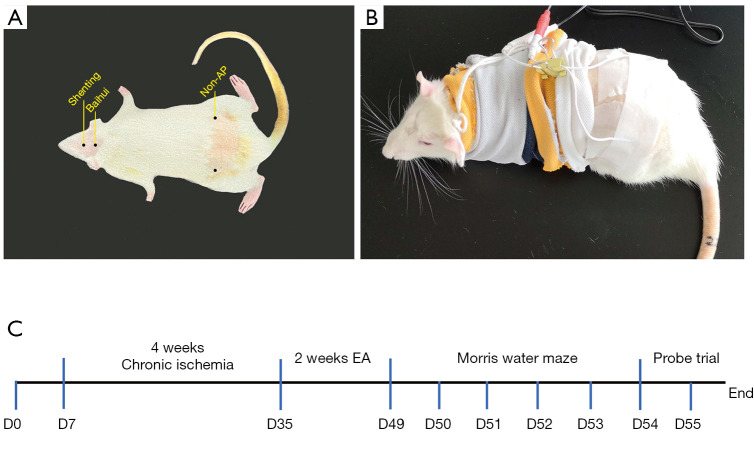
Background: There is currently no effective treatment for vascular dementia (VaD). Scalp electroacupuncture (EA) has served clinically as an alternative treatment for VaD, but its mechanism is still unclear. In this study, we investigated the effect of EA at the Baihui (GV 20) and Shenting (GV 24) acupoints on spatial learning and memory ability, and the expression level of microRNA-81 (miR-81), interleukin-16 (IL-16), and postsynaptic density protein-95 (PSD-95) in the frontal cortex of VaD rats.

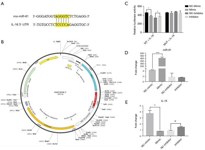
Methods: Male Sprague-Dawley rats were randomly divided into four groups, sham, VaD, non-acupuncture (non-AP) and EA group. The VaD model was established by permanent bilateral occlusion of the common carotid arteries. Morris Water Maze was used to assess the rats' spatial learning and memory. Immunochemistry (IHC), quantitative reverse transcription polymerase chain reaction (qRT-PCR), and western blot analysis were performed to detect the expression level of miR-81, IL-16, and PSD-95. Finally, luciferase assay was used to determine the effect of miR-81 on IL-16 expression in PC12 cells.

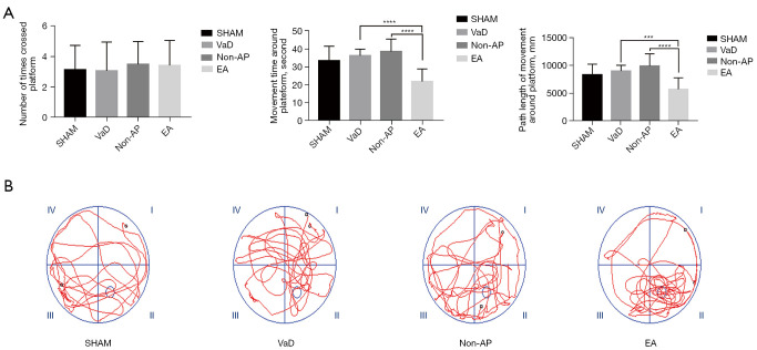
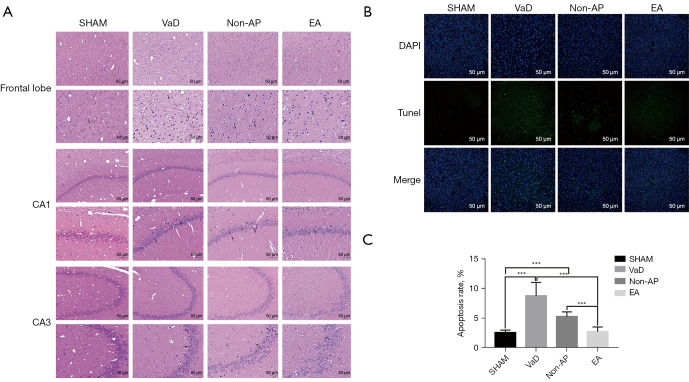
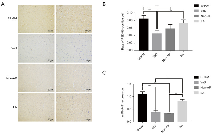
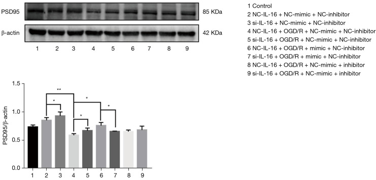
Results: The space exploration experiment of MWM showed the time and distance of the rat's activities around the platform were decreased in the EA group. Compared to the VaD and non-AP group, the number of terminal deoxynucleotidyl transferase-mediated dUDP nick-end labeling (TUNEL)-positive frontal cortical neurons was significantly decreased in EA group. The number of the PSD-95-positive cells and the miR-81 expression level in the frontal cortical in the EA group was dramatically increased in comparison with the other groups. In the PC12 cell validation experiment, IL-16 expression level was reduced under the condition of the miR-81 mimic treatment, while increased in the miR-81 inhibitor group. The PSD-95 protein level was up-regulated in the small interfering (si)RNA-IL16 group compared to the NC-IL16 groups with or without oxygen/glucose deprivation/reperfusion (OGD/R) conditions (P<0.05). However, this was abolished by miR-81 mimic.

Conclusions: In VaD rats, EA may improve spatial learning and memory through miR-81/IL-16/PSD-95 pathway.

Keywords: Baihui acupoint (GV 20); Shenting acupoint (GV 24); electroacupuncture (EA); miR-81; vascular dementia (VaD).


Conflict of interest statement

Conflicts of Interest: All authors have completed the ICMJE uniform disclosure form (available at https://atm.amegroups.com/article/view/10.21037/atm-22-2068/coif). The authors have no conflicts of interest to declare.

Figures

Figure 1
Figure 1
Electroacupuncture rats exhibit recovery of memory (n=12/group). (A) Localization of acupoints and non-acupoints used by electroacupuncture; (B) a self-made rat cover; (C) timeline of animal experiments. EA, electroacupuncture.
Figure 2
Figure 2
Electroacupuncture rats exhibit recovery of memory (n=12/group). (A) Times to cross the platform, path length of movement around the platform, and duration of movement around platform in the four groups during a Morris water maze test, compared with the EA group; (B) the moving trails of rats in the probe trial. VaD, vascular dementia; AP, acupuncture; EA, electroacupuncture. Statistical significance between both groups was defined as ***P<0.001; ****P<0.0001.
Figure 3
Figure 3
Histopathological changed in the frontal lobe, and hippocampal CA1 and CA3 regions (n=6/group). (A) Representative images of morphological changed in cerebral cortex, the hippocampal CA1 and CA3 regions in the sham, VaD, non-AP, and EA groups (n=6/group), 2 weeks after EA staining (HE staining; magnification ×200 and ×400). Scale bar, 50 µm. Nuclear pyknosis was indicated with black arrow. (B) TUNEL staining for apoptosis detection. Expression of apoptotic factors in the cortices of rats among all four groups. Scale bar, 50 µm. (C) Apoptosis rate in the cortex of rats. Data were shown as mean ± SD values, and statistical significance between both groups was defined as ***P<0.001. VaD, vascular dementia; AP, acupuncture; EA, electroacupuncture; HE, hematoxylin and eosin; TUNEL, terminal deoxynucleotidyl transferase-mediated dUDP nick-end labeling; CA1, cornu ammonis 1; CA3, cornu ammonis 3.
Figure 4
Figure 4
The expression of PSD-95 was detected by immunohistochemical staining (n=6/group). (A) Immunohistochemical staining of paraffin sections was used to observe the expression of PSD-95 (n=6/group). Scale bar, 10 µm and 20 µm. (B) The number and grayscale values of PSD-95-positive cells in the EA group were significantly increased compared to the VaD group. Statistical significance between both groups is defined as **P<0.01. ***P<0.001. (C) Expression of miR-81 in cortex of EA group was significantly higher than that of the VaD and non-AP groups. **P<0.01, ***P<0.001. There was no significant difference in miR-81 expression in hippocampal tissue among the groups. VaD, vascular dementia; AP, acupuncture; EA, electroacupuncture; ANOVA, analysis of variance.
Figure 5
Figure 5
The IL-16 3'-UTR is the specific target of miR-81. (A) Binding sites of miR-81 were present in the 3'-UTR of IL-16. TargetScan was used to identify the target sites. (B) Construction profile of the psiCHECK IL-16 plasmid. The binding sequence in the IL-16 3'-UTR was replaced in the psiCHECK-IL-16 MUT plasmid. (C) The relative luciferase activity of psiCHECK-IL-16 was reduced by miR-81. We transfected PC12 cells with the psiCHECK-2-IL-16 vector (200 ng), miR-81 mimic (100 nM), or miR-81 mimic-NC (100 nM) for 48 h using Lipofectamine 2000. *P<0.05 vs. miR-81 NC + psiCHECK-IL-16 group. Data were presented as the mean ± standard error of the mean. Untransfected cells were used as the sham group. (D) The expression of IL-16 mRNA was reduced by miR-81 in PC12 cells. We transfected PC12 cells with miR-81 mimic (100 nM), miR-81 mimic-NC (100 nM), miR-81 inhibitor, and miR-81 inhibitor-NC for 48 h using Lipofectamine 2000. ****P<0.0001. Data were presented as the mean ± standard error of the mean. (E) The expression of IL-16 mRNA was reduced in PC12 cells due to miR-81. **P<0.01 vs. mimic-NC, ##P<0.01 vs. inhibitor-NC. IL-16, interleukin-16; UTR, untranslated region; miR, microRNA; NC, negative control; WT, wild type; MUT, mutant; mRNA, messenger RNA.
Figure 6
Figure 6
Western blot analysis of the role of IL-16 in miR-81 regulating PSD-95 expression. PC12 cells were co-transfected for 48 h with si-IL-16 (50 nM), the NC siRNA (50 nM) and miR-81 mimic (50 nM), miR-81 inhibitor (100 nM), miR-81 mimic-NC (50 nM), miR-81 inhibitor-NC (100 nM) using Lipofectamine 2000. *P<0.05; **P<0.01. Data are presented as the mean ± standard error of the mean. NC, negative control; siRNA, small interfering RNA.

Similar articles

Cited by

References

    1. Korczyn AD, Vakhapova V, Grinberg LT. Vascular dementia. J Neurol Sci 2012;322:2-10. 10.1016/j.jns.2012.03.027 - DOI - PMC - PubMed
    1. Bir SC, Khan MW, Javalkar V, et al. Emerging Concepts in Vascular Dementia: A Review. J Stroke Cerebrovasc Dis 2021;30:105864. 10.1016/j.jstrokecerebrovasdis.2021.105864 - DOI - PubMed
    1. Wang XX, Zhang B, Xia R, et al. Inflammation, apoptosis and autophagy as critical players in vascular dementia. Eur Rev Med Pharmacol Sci 2020;24:9601-14. - PubMed
    1. Rouhl RP, Damoiseaux JG, Lodder J, et al. Vascular inflammation in cerebral small vessel disease. Neurobiol Aging 2012;33:1800-6. 10.1016/j.neurobiolaging.2011.04.008 - DOI - PubMed
    1. Liu X, Zhang R, Wu Z, et al. miR-134-5p/Foxp2/Syn1 is involved in cognitive impairment in an early vascular dementia rat model. Int J Mol Med 2019;44:1729-40. 10.3892/ijmm.2019.4331 - DOI - PMC - PubMed