. 2022 Jul 15;11(7):bio059408.
doi: 10.1242/bio.059408. Epub 2022 Jul 25.

Minimal functional domains of the core polarity regulator Dlg

Affiliations

Minimal functional domains of the core polarity regulator Dlg

Mark J Khoury et al. Biol Open. .

Abstract

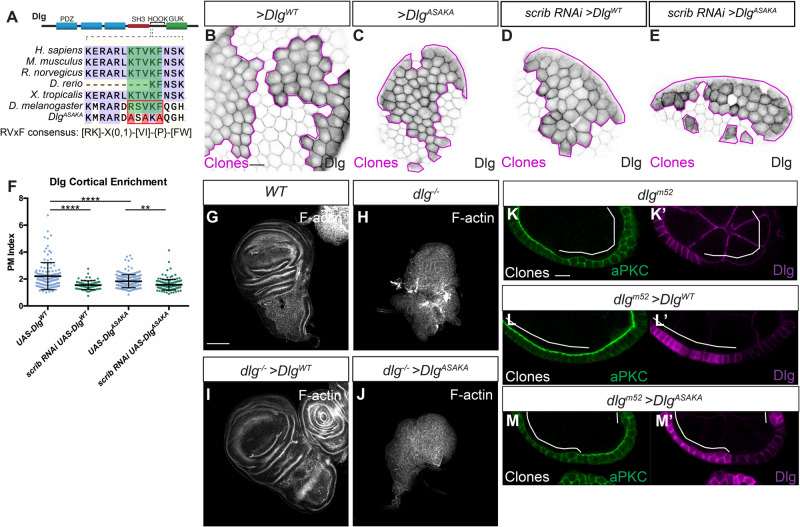
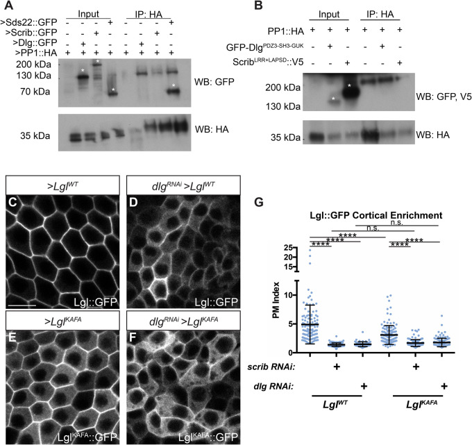
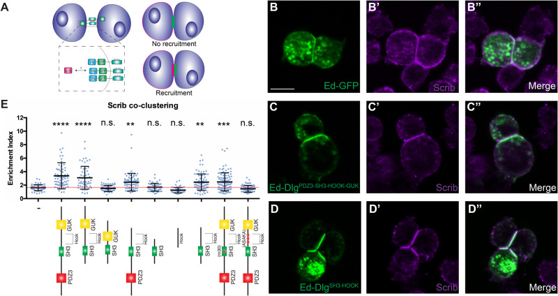
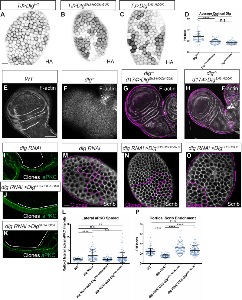
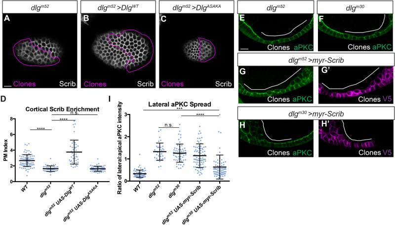
The compartmentalized domains of polarized epithelial cells arise from mutually antagonistic actions between the apical Par complex and the basolateral Scrib module. In Drosophila, the Scrib module proteins Scribble (Scrib) and Discs-large (Dlg) are required to limit Lgl phosphorylation at the basolateral cortex, but how Scrib and Dlg could carry out such a 'protection' activity is not clear. We tested Protein Phosphatase 1α (PP1) as a potential mediator of this activity, but demonstrate that a significant component of Scrib and Dlg regulation of Lgl is PP1 independent, and found no evidence for a Scrib-Dlg-PP1 protein complex. However, the Dlg SH3 domain plays a role in Lgl protection and, in combination with the N-terminal region of the Dlg HOOK domain, in recruitment of Scrib to the membrane. We identify a 'minimal Dlg' comprised of the SH3 and HOOK domains that is both necessary and sufficient for Scrib localization and epithelial polarity function in vivo. This article has an associated First Person interview with the first author of the paper.

Keywords: Cell; Dlg; Polarity; Scrib; Scrib module.


Conflict of interest statement

Competing interests The authors declare no competing or financial interests.

Figures

Fig. 1.
Fig. 1.
The Dlg RVxF motif is critical for function. (A) Cartoon showing location of the RVxF motif in the Dlg protein and conservation of the motif across species. The resides mutated in DlgASAKA are highlighted in red. The RVxF consensus as defined by Wakula et al. (2003) is shown. (B,C) Like DlgWT (B), DlgASAKA localizes to the basolateral membrane and is enriched at the cell cortex (C). (D-F) DlgWT localization (D) as well as DlgASAKA localization (E) is sensitive to scrib depletion, quantified in F. (G,H) Compared to WT (G), dlg null mutant wing discs form disorganized tumors (H). (I,J) Expression of DlgWT rescues this phenotype (I), while expression of DlgASAKA does not rescue (J). (K,L) In the follicle epithelium, dlgm52 null mutants (K) lose polarity, characterized by lateral aPKC spread, and this is rescued by expressing DlgWT (L). (M) In contrast, polarity loss is not rescued by DlgASAKA expression. Scale bars: 10 µm (B,K), 100 µm (G). Magenta lines in B-E and white lines in K-M indicate clones of given genotype. Clones in B-E are flip-out GAL4 clones and those in K-M are MARCM clones. (F) One-way ANOVA with Tukey's multiple comparisons test. Error bars represent s.d. Data points are PM Index measurements in single cells. PM Index=cortical/cytoplasmic intensity. **P<0.01, ****P<0.0001.
Fig. 2.
Fig. 2.
Scrib and Dlg regulate Lgl independently of PP1. (A) Co-IP of transgenic Scrib or Dlg and PP1 from follicle cells fails to detect an interaction, although interaction between PP1 and Sds22 is robustly captured. (B) Co-IP of overexpressed Dlg or Scrib and PP1 from S2 cells following in situ crosslinking also failed to reliably detect interaction between these proteins. Asterisks in A and B indicate relevant bands. (C,D) LglWT membrane localization (C) is severely disrupted by dlg RNAi (D). (E,F) LglKAFA (E) has increased cytoplasmic localization compared to LglWT and is further decreased by dlg RNAi (F). (G) Quantification of Lgl membrane localization. Scale bar: 10 µm. Clones in C-F are flip-out GAL4 clones. (G) One-way ANOVA with Tukey's multiple comparisons test. Error bars represent s.d. PM Index=cortical/cytoplasmic intensity. Data points are measurements from individual cells. n.s. (not significant) P>0.05, ****P<0.0001.
Fig. 3.
Fig. 3.
DlgSH3-HOOK is sufficient for Scrib localization in an induced polarity system. (A) Cartoon of S2 induced polarity assay. Polarizing Dlg by fusion to Ed enables testing of Scrib recruitment in a minimal synthetic system. (B) S2 cells expressing Ed-GFP can be clustered by adhesion between Ed molecules, but this does not alter Scrib localization. (C) When DlgPDZ3-SH3-HOOK-GUK is fused to Ed, it creates a polarity crescent at the contact site that is able to recruit Scrib. (D) A minimal fragment, DlgSH3-HOOK retains the ability to recruit Scrib to the contact site. (E) Quantification of Scrib recruitment to the polarity site in various Ed-Dlg constructs schematized below. DlgPDZ3-SH3-HOOK-GUK is able to enrich Scrib, while the Ed-GFP negative control cannot. The HOOK and SH3 domains are necessary and, when in combination, sufficient to recruit Scrib. Statistical tests are versus the Ed-GFP negative control construct. Red line denotes the average for the Ed-GFP negative control and indicates no Scrib contact site enrichment. Scale bar: 10 µm. (E) One-way ANOVA with Tukey's multiple comparisons test. Error bars indicate s.d. Data points are individual cell clusters. Enrichment index=contact site/non-contact site intensity. n.s. (not significant) P>0.05, **P<0.01, ***P<0.001, ****P<0.0001.
Fig. 4.
Fig. 4.
Dlg SH3 and HOOK domains are sufficient for function in vivo. (A-C) Like WT Dlg (A), DlgSH3-HOOK-GUK (B) and DlgSH3-HOOK (C) localize to the basolateral membrane in follicle cells. All constructs contain an HA epitope tag used for detection. (D) Quantification of cortical localization in A-C. (E-H) Compared to WT (E) and dlg null mutants (F), DlgSH3-HOOK-GUK (G) and DlgSH3-HOOK (H) fully rescue polarity and epithelial architecture in wing imaginal discs. (I-L) In monolayered dlg-depleted follicle cells (I), both DlgSH3-HOOK-GUK (J) and DlgSH3-HOOK (K) provide polarity-rescuing activity, quantitated in L. Full restoration of monolayering is more efficient by DlgSH3-HOOK-GUK than DlgSH3-HOOK: 89.5% (n=38) of DlgSH3-HOOK-GUK show no regions of multilayering in rescued follicles, compared to 20.5% (n=39) of DlgSH3-HOOK rescued follicles and 0% (n=37) of follicles with dlg-depleted clones alone. (M-P) Both DlgSH3-HOOK-GUK (N) and DlgSH3-HOOK (O) fully rescue loss of cortical Scrib seen in dlg-depleted cells (M), quantified in P. Scale bars: 10 µm (A,I,M), 100 µm (E). White or magenta lines indicate clones of given genotypes; Clones in I-K and M-O are flip-out GAL4 clones. (D,L,P) One-way ANOVA with Tukey's multiple comparisons test. Error bars indicate s.d. PM Index=cortical/cytoplasmic intensity. aPKC spread is a ratio of lateral:apical fluorescence intensity. Data points are individual cell measurements. n.s. (not significant) P>0.05, **P<0.01, ***P<0.001, ****P<0.0001.
Fig. 5.
Fig. 5.
Dlg SH3-HOOK is primarily required to regulate Scrib localization. (A) dlgm52 null mutant cells show reduced cortical localization of Scrib. (B) Scrib mislocalization is rescued by expression of DlgWT. (C) Scrib mislocalization is not rescued by expression of DlgASAKA. (D) Quantification of Scrib localization in A-C. (E,F) Both dlgm52 null mutants (E) and dlgm30 SH3 point mutant cells (F) lose polarity and mislocalize aPKC. (G,H) Preventing Scrib mislocalization by cortical tethering (myr-Scrib) partially suppresses the polarity loss phenotypes of dlgm30 SH3 mutant cells (H) but not dlgm52 null mutant cells (G). (G′,H′) myr-Scrib contains a V5 epitope tag used for detection. (I) Quantification of aPKC mislocalization in (E-H). Scale bars: 10 µm. Magenta or white lines indicate MARCM clones of given genotypes. (D,I) One-way ANOVA with Tukey's multiple comparisons test. Error bars indicate s.d. PM Index=cortical/cytoplasmic intensity. aPKC spread is a ratio of lateral:apical fluorescence intensity. Data points are individual cell measurements. n.s. (not significant) P>0.05, ***P<0.001, ****P<0.0001.

Similar articles

Cited by

References

    1. Albertson, R., Chabu, C., Sheehan, A. and Doe, C. Q. (2004). Scribble protein domain mapping reveals a multistep localization mechanism and domains necessary for establishing cortical polarity. J. Cell Sci. 117, 6061-6070. 10.1242/jcs.01525 - DOI - PubMed
    1. Awadia, S., Huq, F., Arnold, T. R., Goicoechea, S. M., Sun, Y. J., Hou, T., Kreider-Letterman, G., Massimi, P., Banks, L., Fuentes, E. J.et al. (2019). SGEF forms a complex with Scribble and Dlg1 and regulates epithelial junctions and contractility. J. Cell Biol. 218, 2699-2725. 10.1083/jcb.201811114 - DOI - PMC - PubMed
    1. Bailey, M. J. and Prehoda, K. E. (2015). Establishment of Par-polarized cortical domains via phosphoregulated membrane motifs. Dev. Cell 35, 199-210. 10.1016/j.devcel.2015.09.016 - DOI - PMC - PubMed
    1. Bellaïche, Y., Radovic, A., Woods, D. F., Hough, C. D., Parmentier, M.-L., O'Kane, C. J., Bryant, P. J. and Schweisguth, F. (2001). The partner of inscuteable/discs-large complex is required to establish planar polarity during asymmetric cell division in Drosophila. Cell 106, 355-366. 10.1016/S0092-8674(01)00444-5 - DOI - PubMed
    1. Bergstralh, D. T., Lovegrove, H. E. and St Johnston, D. (2013). Discs large links spindle orientation to apical-basal polarity in drosophila epithelia. Curr. Biol. 23, 1707-1712. 10.1016/j.cub.2013.07.017 - DOI - PMC - PubMed

Publication types

Substances