Observational Study
. 2022 Aug 2;146(5):383-397.
doi: 10.1161/CIRCULATIONAHA.121.057145. Epub 2022 Jun 20.

Cerebral Microbleeds During Transcatheter Aortic Valve Replacement: A Prospective Magnetic Resonance Imaging Cohort

Affiliations
Observational Study

Cerebral Microbleeds During Transcatheter Aortic Valve Replacement: A Prospective Magnetic Resonance Imaging Cohort

Eric Van Belle et al. Circulation. .

Abstract

Background: Cerebral microbleeds (CMBs) have been observed in healthy elderly people undergoing systematic brain magnetic resonance imaging. The potential role of acute triggers on the appearance of CMBs remains unknown. We aimed to describe the incidence of new CMBs after transcatheter aortic valve replacement (TAVR) and to identify clinical and procedural factors associated with new CMBs including hemostatic measures and anticoagulation management.

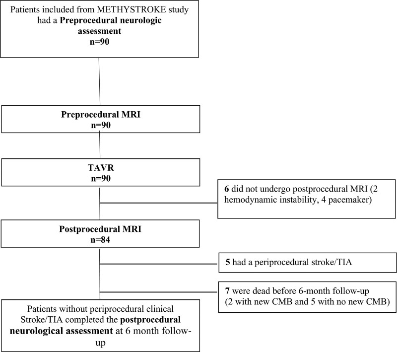
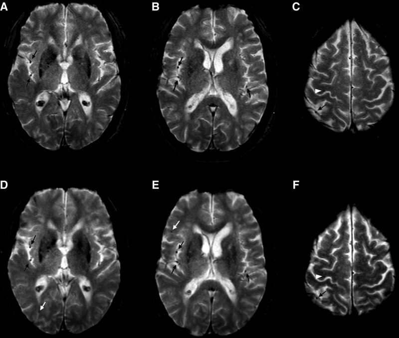
Methods: We evaluated a prospective cohort of patients with symptomatic aortic stenosis referred for TAVR for CMBs (METHYSTROKE [Identification of Epigenetic Risk Factors for Ischemic Complication During the TAVR Procedure in the Elderly]). Standardized neurologic assessment, brain magnetic resonance imaging, and analysis of hemostatic measures including von Willebrand factor were performed before and after TAVR. Numbers and location of microbleeds on preprocedural magnetic resonance imaging and of new microbleeds on postprocedural magnetic resonance imaging were reported by 2 independent neuroradiologists blinded to clinical data. Measures associated with new microbleeds and postprocedural outcome including neurologic functional outcome at 6 months were also examined.

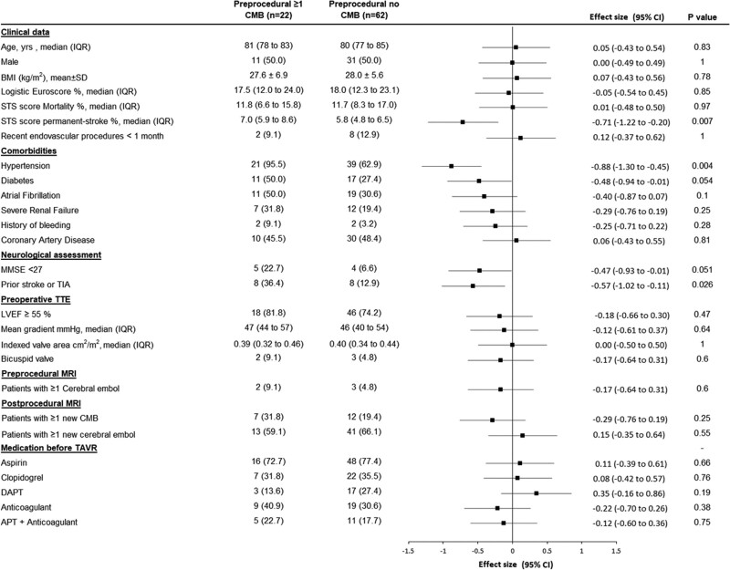
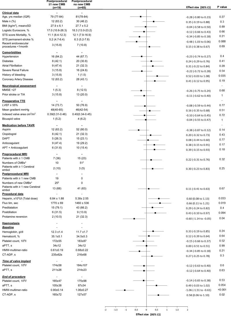
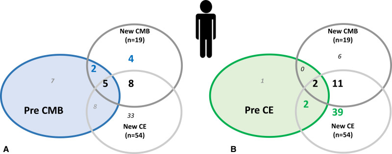
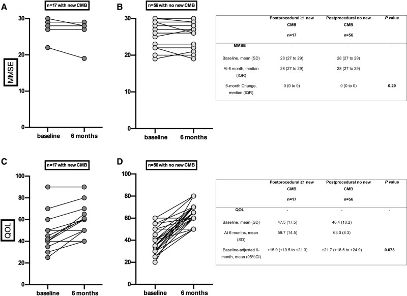
Results: A total of 84 patients (47% men, 80.9±5.7 years of age) were included. On preprocedural magnetic resonance imaging, 22 patients (26% [95% CI, 17%-37%]) had at least 1 microbleed. After TAVR, new microbleeds were observed in 19 (23% [95% CI, 14%-33%]) patients. The occurrence of new microbleeds was independent of the presence of microbleeds at baseline and of diffusion-weighted imaging hypersignals. In univariable analysis, a previous history of bleeding (P=0.01), a higher total dose of heparin (P=0.02), a prolonged procedure (P=0.03), absence of protamine reversion (P=0.04), higher final activated partial thromboplastin time (P=0.05), lower final von Willebrand factor high-molecular-weight:multimer ratio (P=0.007), and lower final closure time with adenosine-diphosphate (P=0.02) were associated with the occurrence of new postprocedural microbleeds. In multivariable analysis, a prolonged procedure (odds ratio, 1.22 [95% CI, 1.03-1.73] for every 5 minutes of fluoroscopy time; P=0.02) and postprocedural acquired von Willebrand factor defect (odds ratio, 1.42 [95% CI, 1.08-1.89] for every lower 0.1 unit of high-molecular-weight:multimer ratio; P=0.004) were independently associated with the occurrence of new postprocedural microbleeds. New CMBs were not associated with changes in neurologic functional outcome or quality of life at 6 months.

Conclusions: One out of 4 patients undergoing TAVR has CMBs before the procedure and 1 out of 4 patients develops new CMBs. Procedural or antithrombotic management and persistence of acquired von Willebrand factor defect were associated with the occurrence of new CMBs.

Registration: URL: https://www.

Clinicaltrials: gov; Unique identifier: NCT02972008.

Keywords: aortic valve stenosis; cerebral microbleeds; hemostasis; transcatheter aortic valve replacement; von Willebrand factor.


Figures

Figure 1.
Figure 1.
Flowchart of clinical and imaging evaluations. CMB indicates cerebral microbleed; MRI, magnetic resonance imaging; TAVR, transcatheter aortic valve implantation; and TIA, transient ischemic attack.
Figure 2.
Figure 2.
T2*-weighted gradient-echo sequence before and after transcatheter aortic valve implantation. Before (A–C) and after (D–F) transcatheter aortic valve implantation. This patient had a unique microbleed in the right precentral gyrus before transcatheter aortic valve implantation (white arrowhead; C). Two new microbleeds were observed after transcatheter aortic valve implantation, in the right frontal and temporal lobes (white arrows; D and E). Cerebral microbleeds were identified as homogeneous, round foci, <10 mm diameter, of low signal intensity. Flow void artifacts of the pial blood vessels (black arrows) were clearly distinguished from cerebral microbleeds by their location in the subarachnoid space and their tubular morphology on the adjacent slices. Preoperative and postoperative T2* images have been coregistered with an automated coregistration software dedicated to longitudinal comparison of magnetic resonance images using rigid transformations on the basis of mutual information (Longitudinal Brain Imaging; Philips Medical Systems).
Figure 3.
Figure 3.
Baseline factors associated with the presence of cerebral microbleeds on preprocedural magnetic resonance imaging. Data are mean (SD), median (interquartile range [IQR]), or n (%) unless otherwise indicated. P values were calculated using the Fisher exact test for categorical variables or the Student t test (or the Mann-Whitney U test for non-Gaussian distribution) for quantitative variables. Effect sizes were the standardized differences (calculated on rank-transformed values for non-Gaussian quantitative variables); absolute values of 0.2, 0.5, and 0.8 were interpreted as small, medium, and large effect size. APT indicates antiplatelet therapy; BMI, body mass index; DAPT, dual antiplatelet therapy; LVEF, left ventricular ejection fraction; MMSE, Mini-Mental State Examination; MRI, magnetic resonance imaging; STS, Society of Thoracic Surgeons; TAVR, transcatheter aortic valve replacement; TIA, transient ischemic attack; and TTE, transthoracic echography.
Figure 4.
Figure 4.
Baseline and procedural factors associated with new cerebral microbleeds. Data are mean (SD), median (interquartile range [IQR]), or n (%) unless otherwise indicated. New postprocedural cerebral microbleeds (CMBs) are CMBs present on postprocedural magnetic resonance imaging (MRI) that were not present on the preprocedural MRI. P values were calculated using the Fisher exact test for categorical variables or the Student t test (or the Mann-Whitney U test for non-Gaussian distribution) for quantitative variables. Effect sizes were the standardized differences (calculated on rank-transformed values for non-Gaussian quantitative variables); absolute values of 0.2, 0.5, and 0.8 were interpreted as small, medium, and large effect size. *Numbers of CMBs in the whole (sub)group. †One patient had 31 CMBs as part of a severe brain microangiopathy. ‡All 25 new CMBs were ≤5 mm. APT indicates antiplatelet therapy; aPTT, activated partial thromboplastin time; BMI, body mass index; CT-ADP, closure time with adenosine diphosphate (refers to platelet function analyzer); DAPT, dual antiplatelet therapy; HMW, high molecular weight; LVEF, left ventricular ejection fraction; MMSE, Mini-Mental State Examination; STS, Society of Thoracic Surgeons; TAVR, transcatheter aortic valve replacement; TIA, transient ischemic attack; and TTE, transthoracic echography.
Figure 5.
Figure 5.
Repartition of patients according to the presence of new CMB alone, new CE alone, or both on postprocedural MRI according to the presence of CMB or CE on preprocedural MRI. Repartition of patients according to the presence of new cerebral microbleed (CMB) alone (n=6), new cerebral embol (CE) alone (n=41), or both (n=13) on postprocedural magnetic resonance imaging (MRI) according to the presence of CMB on preprocedural MRI (A) and the presence of CE on preprocedural MRI (B). Numbers in the colored circles represent the number of patients presenting with previous CMB (A) or CE (B) alone or associated with new CMB or new CE alone or in association. No relation was found between new postprocedural microbleed and preprocedural microbleed (P=0.23), preprocedural CE (P=0.33), or new postprocedural CE (P=0.88).
Figure 6.
Figure 6.
Neurologic and quality of life evolution from before the procedure to 6-month follow-up according to the occurrence of new cerebral microbleeds after TAVR in the 73 patients without periprocedural stroke or TIA who were alive at 6 months. Cognition assessed by Mini-Mental State Examination (MMSE) score at baseline and after 6 months in the presence (A) or absence (B) of new cerebral microbleeds (CMBs). Quality of life (QOL) assessed by EQ-5D score at baseline and after 6 months in the presence (C) or absence (D) of new CMBs. Patients with periprocedural stroke or transient ischemic attack (TIA) or who were deceased at 6 months were excluded. Baseline-adjusted effect sizes were standardized differences (calculated on rank-transformed values for MMSE). P value calculated using analysis of covariance on 6-month change adjusted for baseline values (calculated on rank-transformed values for MMSE).

References

    1. Greenberg SM, Vernooij MW, Cordonnier C, Viswanathan A, Al-Shahi Salman R, Warach S, Launer LJ, Van Buchem MA, Breteler MM, Microbleed Study Group. Cerebral microbleeds: a guide to detection and interpretation. Lancet Neurol. 2009;8:165–174. doi: 10.1016/S1474-4422 (09)70013-4 - PMC - PubMed
    1. Cordonnier C, Demchuk A, Ziai W, Anderson CS. Intracerebral haemorrhage: current approaches to acute management. Lancet. 2018;392:1257–1268. doi: 10.1016/S0140-6736(18)31878-6 - PubMed
    1. Cordonnier C, Potter GM, Jackson CA, Doubal F, Keir S, Sudlow CLM, Wardlaw JM, Al-Shahi Salman R. Improving interrater agreement about brain microbleeds: development of the Brain Observer MicroBleed Scale (BOMBS). Stroke J Cereb Circ. 2009;40:94–99. doi: 10.1161/STROKEAHA.108.526996 - PubMed
    1. Haller S, Vernooij MW, Kuijer JPA, Larsson E-M, Jäger HR, Barkhof F. Cerebral microbleeds: imaging and clinical significance. Radiology. 2018;287:11–28. doi: 10.1148/radiol.2018170803 - PubMed
    1. Wardlaw JM, Smith EE, Biessels GJ, Cordonnier C, Fazekas F, Frayne R, Lindley RI, O’Brien JT, Barkhof F, Benavente OR, et al. . Neuroimaging standards for research into small vessel disease and its contribution to ageing and neurodegeneration. Lancet Neurol. 2013;12:822–838. doi: 10.1016/S1474-4422 (13)70124-8 - PMC - PubMed

Publication types

Associated data