

Neuroimaging Correlates of Cognitive Deficits in Wilson's Disease
- PMID: 35723521
- PMCID: PMC9542291
- DOI: 10.1002/mds.29123
Neuroimaging Correlates of Cognitive Deficits in Wilson's Disease
Abstract
Background: Cognitive impairment is common in neurological presentations of Wilson's disease (WD). Various domains can be affected, and subclinical deficits have been reported in patients with hepatic presentations. Associations with imaging abnormalities have not been systematically tested.
Objective: The aim was to determine the neuroanatomical basis for cognitive deficits in WD.
Methods: We performed a 16-item neuropsychological test battery and magnetic resonance brain imaging in 40 patients with WD. The scores for each test were compared between patients with neurological and hepatic presentations and with normative data. Associations with Unified Wilson's Disease Rating Scale neurological examination subscores were examined. Quantitative, whole-brain, multimodal imaging analyses were used to identify associations with neuroimaging abnormalities in chronically treated stable patients.
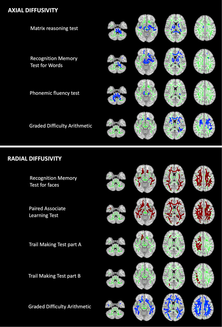
Results: Abstract reasoning, executive function, processing speed, calculation, and visuospatial function scores were lower in patients with neurological presentations than in those with hepatic presentations and correlated with neurological examination subscores. Deficits in abstract reasoning and phonemic fluency were associated with lower putamen volumes even after controlling for neurological severity. About half of patients with hepatic presentations had poor performance in memory for faces, cognitive flexibility, or associative learning relative to normative data. These deficits were associated with widespread cortical atrophy and/or white matter diffusion abnormalities.
Conclusions: Subtle cognitive deficits in patients with seemingly hepatic presentations represent a distinct neurological phenotype associated with diffuse cortical and white matter pathology. This may precede the classical neurological phenotype characterized by movement disorders and executive dysfunction and be associated with basal ganglia damage. A binary phenotypic classification for WD may no longer be appropriate. © 2022 The Authors. Movement Disorders published by Wiley Periodicals LLC on behalf of International Parkinson and Movement Disorder Society.
Keywords: Wilson's disease; cognition; magnetic resonance imaging.
© 2022 The Authors. Movement Disorders published by Wiley Periodicals LLC on behalf of International Parkinson and Movement Disorder Society.
Figures


References
-
- Frota NA, Barbosa ER, Porto CS, et al. Cognitive impairment and magnetic resonance imaging correlations in Wilson's disease. Acta Neurol Scand 2013;127(6):391–398. - PubMed
-
- Seniów J, Bak T, Gajda J, et al. Cognitive functioning in neurologically symptomatic and asymptomatic forms of Wilson's disease. Mov Disord 2002;17(5):1077–1083. - PubMed
-
- Lang C, Müller D, Claus D, et al. Neuropsychological findings in treated Wilson's disease. Acta Neurol Scand 1990;81(1):75–81. - PubMed
-
- CA K, AG B. Psychological impairment in Wilson's disease. J Nerv Ment Dis 1956;124(3):251–255. - PubMed
Publication types
MeSH terms
Grants and funding
LinkOut - more resources
Full Text Sources
Medical
