. 2022 Dec 16;130(6):785-798.
doi: 10.1093/aob/mcac083.

A protoxylem pathway to evolution of pith? An hypothesis based on the Early Devonian euphyllophyte Leptocentroxyla

Affiliations

A protoxylem pathway to evolution of pith? An hypothesis based on the Early Devonian euphyllophyte Leptocentroxyla

Alexandru M F Tomescu et al. Ann Bot. .

Abstract

Background and aims: The Early Devonian (Emsian, 400-395 Ma) tracheophyte Leptocentroxyla tetrarcha Bickner et Tomescu emend. Tomescu et McQueen combines plesiomorphic Psilophyton-type tracheid thickenings with xylem architecture intermediate between the plesiomorphic basal euphyllophyte haplosteles and the complex actinosteles of Middle Devonian euphyllophytes. We document xylem development in Leptocentroxyla based on anatomy and explore its implications, which may provide a window into the evolution of pith.

Methods: Leptocentroxyla is preserved by permineralization in the Battery Point Formation (Quebec, Canada). Serial sections obtained using the cellulose acetate peel technique document branching pattern, anatomy of trace divergence to appendages, protoxylem architecture, and variations in tracheid size and wall thickening patterns.

Key results: Leptocentroxyla has opposite decussate pseudo-whorled branching and mesarch protoxylem, and represents the earliest instance of central histological differentiation in a euphyllophyte actinostele. Tracheids at the centre of xylem exhibit simplified Psilophyton-type wall thickenings and are similar in size (at the axis centre) or smaller than the surrounding metaxylem tracheids (at the centre of appendage traces).

Conclusions: The position and developmental attributes of the simplified Psilophyton-type tracheids suggest they may have been generated by the protoxylem developmental pathway. This supports the delayed and shortened protoxylem differentiation hypothesis, which explains the evolution of pith by (1) delay in the onset of differentiation and lengthening of cell growth duration in a central protoxylem strand; and (2) shortening of the interval of differentiation of those tracheids, leading to progressive simplification (and eventual loss) of secondary wall thickenings, and replacement of tracheids with a central parenchymatous area. NAC domain transcription factors and their interactions with abscisic acid may have provided the regulatory substrate for the developmental changes that led to the evolution of pith. These could have been orchestrated by selective pressures associated with the expansion of early vascular plants into water-stresses upland environments.

Keywords: Leptocentroxyla; Anatomy; Devonian; euphyllophyte; evo-devo; fossil; pith; protoxylem; tracheary element differentiation; xylem development.


Figures

Fig. 1.
Fig. 1.
Leptocentroxyla tetrarcha, axis preservation. (A) Position of the axis in two slabs that were sectioned using the cellulose acetate peel technique; letters indicate the approximate position of the sections illustrated in B–E. Note variation in the quality of preservation along the axis, from good at the proximal end (E) to more distorted at the distal end (B). (B) 557820-3 Atop 186f. (C) 557820-3 Abot 2d. (D) 557820-3 Btop 150b. (E) 557820-3 Bbot 90f. Scale bar = 500 µm.
Fig. 2.
Fig. 2.
Leptocentroxyla tetrarcha, anatomy of trace divergence to lateral appendages, protoxylem architecture and tracheid pitting. Serial sections (A–C, basal to distal) of a xylem trace pinching off from the tip of a xylem lobe (A, B) and diverging in radial direction through the inner cortex (B, C); note radially elongate shape of trace to lateral appendage and groups of sclerenchyma in the cortex (on right side of xylem lobe and appendage trace). Groups of three or four smaller metaxylem tracheids close to the tips of the xylem lobe indicate the position of inconspicuous protoxylem strands (arrowheads in A–E); note that although protoxylem tracheids are not always recognizable, the patterning of tracheid sizes indicates the position of protoxylem strands. Distally to the trace divergence point, a second protoxylem strand is present (arrowheads in D and E), interpreted as resulting from a divergence in the single protoxylem strand at the lobe tip in C. The metaxylem of the main axis (F, G) features Psilophyton-type thickenings (F and G top and bottom), except for the central tracheids (G, centre and H), which are thinner-walled and characterized by scalariform wall thickenings that separate horizontally elongated oval bordered pits; note remnants of rare partitions separating the large apertures of these bordered pits into smaller apertures (arrowheads in H). (A) 557820-3 Bbot 87f. (B) 557820-3 Bbot 90f. (C) 557820-3 Bbot 190f. (D) 557820-3 Bbot 119f. (E) 557820-3 Bbot 182f. (F-H). 557820-3 Bbot 110. Scale bars = 150 µm (A–E); 50 µm (F, G); 35 µm (H).
Fig. 3.
Fig. 3.
Leptocentroxyla tetrarcha, branching architecture as reflected by the pattern of trace emission to lateral appendages. A series of three cross-sections (C proximal to A distal) through a branching region demonstrates pseudo-whorled branching. Whereas one pair of opposite traces that are pinching off in C have already separated from the stele in B (orange arrowheads; bottom right part of the xylem in C is distorted taphonomically), the other pair of opposite traces (black arrowheads) are just pinching off throughout the sequence (C–B–A) and are separating at a level distal to the topmost section (A). Note missing trace at bottom right in A (asterisk), which has exited the axis and entered the base of a branch at this level. The inset diagram shows the position of the three planes of section (orange lines corresponding, from top to bottom, to A, B and C) with respect to the branching of the xylem (grey central band) and the axis; the darker dots in the xylem represent traces of appendages diverging perpendicular to the plane of the diagram. The divergence of the two pairs of opposite traces is only slightly offset vertically, which renders the branching pattern similar to a whorled arrangement. (A) 557820-3 Bbot 159f. (B) 557820-3 Bbot 119f. (C) 557820-3 Bbot 87f. Scale bar = 500 µm.
Fig. 4.
Fig. 4.
Leptocentroxyla tetrarcha, secondary wall thickenings of tracheids in lateral appendages. The tracheids in the traces to lateral appendages feature Psilophyton-type thickenings (A, B), except for the central region of the trace, which consists of smaller tracheids with thinner walls characterized by scalariform thickenings that separate oval horizontal bordered pits (A, centre; B, centre; C); note pit in C (centre, arrowhead) with aperture separated by a median vertical partition and remnant of such a partition in a pit in B (arrowhead). (A) 557820-3 Bbot 90f. Scale bar = 50 µm. (B) 557820-3 Bbot 110. Scale bar = 30 µm. (C) 557820-3 Bbot 90f. Scale bar = 25 µm.
Fig. 5.
Fig. 5.
Leptocentroxyla tetrarcha, tracheid size variation. (A) Location of the central (purple), peripheral (green) and lobe areas (blue) of the axis xylem where maximum tracheid diameters were measured in each section. 557820-3 Bbot 90f. Scale bar = 200 µm. (B) Maximum metaxylem tracheid diameters in the central, peripheral and lobe tracheids (N = 40 for each tracheid type); bottom whisker = 25th percentile − 1.5 × inter-quartile range; top whisker = 75th percentile + 1.5 × inter-quartile range; dots = outliers; horizontal line = median; dashed horizontal line = mean. (C) Maximum metaxylem tracheid size is (weakly) positively correlated with the number of thin-walled tracheids in the central area of the xylem (r = 0.296, central tracheids; r = 0.376, peripheral tracheids; r = 0.290, lobe tracheids).
Fig. 6.
Fig. 6.
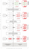
A possible path to the evolution of pith as explained by the delayed and shortened protoxylem differentiation hypothesis – by central histological differentiation in a protostele; grey vertical lines and cylinder tops/bases denote immature and differentiating cells; coloured vertical lines and cylinder tops/bases denote mature tissues: red = tracheids, green = parenchyma. The starting point is a euphyllophyte with centrarch primary xylem maturation (i.e. a central protoxylem strand), like the Early Silurian Psilophyton, wherein tracheids of the central protoxylem strand mature (depicted by red secondary wall thickenings) in the zone of cell elongation, whereas the rest of the primary xylem (metaxylem) matures beyond the zone of elongation with production of P-type secondary wall thickenings. From this starting point, delayed differentiation (maturation) of the central tracheids allows them to grow in size before they differentiate; if the delay in differentiation lasts until these cells exit the zone of elongation, then these cells will differentiate in the same way as the metaxylem tracheids, with P-type thickenings (theoretical stage). If the interval of maturation is shortened in the central cells with delayed maturation, these cells will have less time to build secondary wall thickenings, which will result in simpler thickenings, like the central simplified P-type tracheids of Leptocentroxyla. Further shortening of the maturation interval of the central cells of the stele would eventually lead to complete elimination of the secondary wall building phase in the differentiation process of these cells, which will mature into parenchyma cells (as seen in some Devonian moniliformopsids – e.g. Arachnoxylon – and radiatopsids – e.g. Actinoxylon).

Comment in

References

    1. Banks HP, Leclercq S, Hueber FM.. 1975. Anatomy and morphology of Psilophyton dawsonii, sp. n. from the Late Lower Devonian of Quebec (Gaspe), and Ontario, Canada. Palaeontographica Americana 8: 75–127.
    1. Beck CB. 1976. Current status of the Progymnospermopsida. Review of Palaeobotany and Palynology 21: 5–23.
    1. Beck CB, Schmid R, Rothwell GW.. 1982. Stelar morphology and the primary vascular system of seed plants. Botanical Review 48: 691–815.
    1. Beck CB, Galtier J, Stein WE.. 1992. A reinvestigation of Diichnia Read from the New Albany Shale of Kentucky. Review of Palaeobotany and Palynology 75: 1–32.
    1. Beck CB, Stein WE.. 1993. Crossia virginiana gen. et sp. nov., a new member of the Stenokoleales from the Middle Devonian of southwestern Virginia. Palaeontographica B 229: 115–134.

Publication types