. 2022 Jun 20;12(1):10333.
doi: 10.1038/s41598-022-13569-4.

Germline variants of ATG7 in familial cholangiocarcinoma alter autophagy and p62

Affiliations

Germline variants of ATG7 in familial cholangiocarcinoma alter autophagy and p62

Stephanie U Greer et al. Sci Rep. .

Abstract

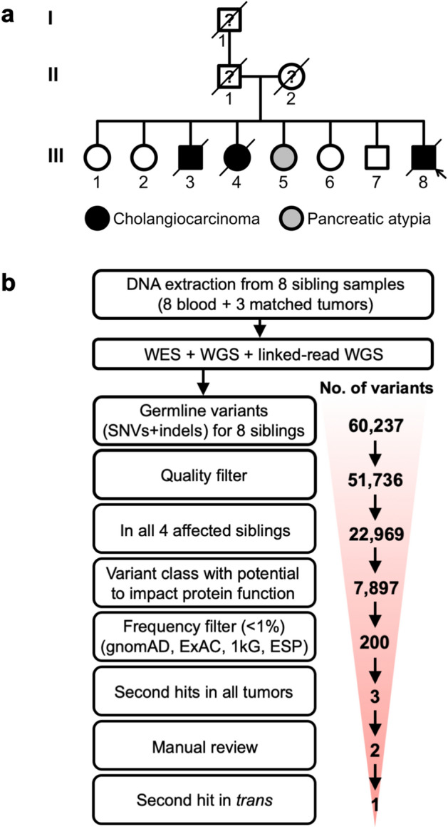
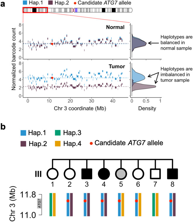
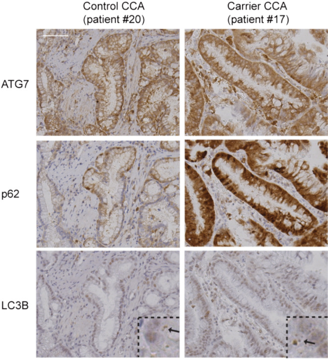
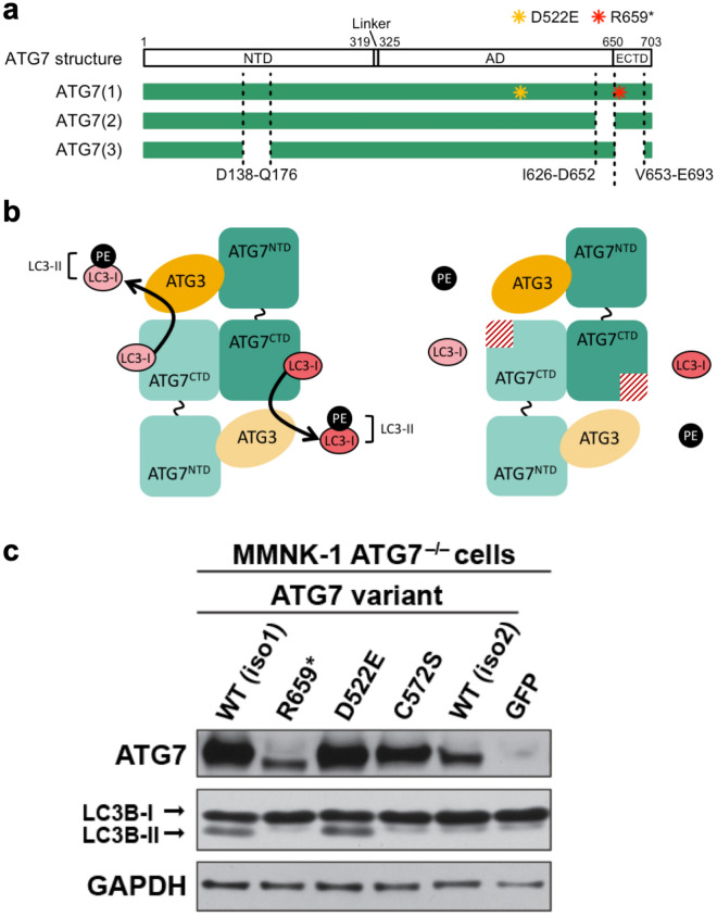
Autophagy is a housekeeping mechanism tasked with eliminating misfolded proteins and damaged organelles to maintain cellular homeostasis. Autophagy deficiency results in increased oxidative stress, DNA damage and chronic cellular injury. Among the core genes in the autophagy machinery, ATG7 is required for autophagy initiation and autophagosome formation. Based on the analysis of an extended pedigree of familial cholangiocarcinoma, we determined that all affected family members had a novel germline mutation (c.2000C>T p.Arg659* (p.R659*)) in ATG7. Somatic deletions of ATG7 were identified in the tumors of affected individuals. We applied linked-read sequencing to one tumor sample and demonstrated that the ATG7 somatic deletion and germline mutation were located on distinct alleles, resulting in two hits to ATG7. From a parallel population genetic study, we identified a germline polymorphism of ATG7 (c.1591C>G p.Asp522Glu (p.D522E)) associated with increased risk of cholangiocarcinoma. To characterize the impact of these germline ATG7 variants on autophagy activity, we developed an ATG7-null cell line derived from the human bile duct. The mutant p.R659* ATG7 protein lacked the ability to lipidate its LC3 substrate, leading to complete loss of autophagy and increased p62 levels. Our findings indicate that germline ATG7 variants have the potential to impact autophagy function with implications for cholangiocarcinoma development.


Conflict of interest statement

The authors declare no competing interests.

Figures

Figure 1
Figure 1
Identification of a putative germline predisposition variant in a family with inherited CCA. (a) This pedigree depicts a family with an inherited predisposition to biliary cancers. The current study had access to samples from generation III of the family. (b) The number of putative germline variants (single nucleotide variants and insertion-deletions) is indicated for each stage of filtering.
Figure 2
Figure 2
Haplotype analysis of the candidate ATG7 allele (p.R659*). (a) Extended haplotype of the 45 Mb deleted region of chromosome 3p in individual III:8. The blocks indicate the original fragmented haplotypes, and their color denotes their subsequent assignment to haplotypes covering many Megabases. The candidate ATG7 allele exists in haplotype 1 (blue), which was the non-deleted haplotype in the tumor of this individual. (b) Germline haplotype analysis of siblings for the ATG7 genomic region. The haplotype segregation across all eight siblings in generation III was determined for the 0.8 Mb genomic region surrounding the ATG7 allele (red dot). The candidate ATG7 allele exists in haplotype 1 (blue).
Figure 3
Figure 3
Increased p62 expression in tumors of ATG7 variant (p.D522E) carriers. The microscopic images show sections of CCA from an rs146589465 (p.D522E) carrier (patient #17) and a non-carrier control (patient #20) stained with antibodies against ATG7, p62 and LC3B. For the LC3B staining, a higher magnification window is shown and the arrows point to LC3 dots that are indicative of autophagosomes. Scale bar represents 20 µm and applies to all panels.
Figure 4
Figure 4
ATG7 p.R659* is a loss-of-function mutation. (a) The predicted structure of ATG7 contains an N-terminal domain (NTD), linker region, adenylation domain (AD), and extreme C-terminal domain (ECTD). The p.R659* (R659*) and p.D522E (D522E) mutations are indicated in isoform 1 of the ATG7 protein. (b) Predicted functional impact of ATG7 p.R659* mutation. Wildtype ATG7 and ATG3 conjugate ATG8 proteins such as LC3 with phosphatidylethanolamine (PE). We determined that the ATG7 R659* mutant lacks PE conjugation activity to LC3 as a result of the ECTD truncation. (c) Expression of ATG7 WT isoform 1 (iso1) and D522E restored the lipidation of LC3B whereas expression of ATG7 R659*, C572S, or WT isoform 2 (iso2) failed to lipidate LC3B from LC3B-I to LC3B-II. A Myc-tag was used to detect the expression of exogenous ATG7 from various vectors. A GFP expressing vector was used as a negative control. GAPDH was used as the loading control. Original blots are presented in Supplementary Fig. S9 online.
Figure 5
Figure 5
Impaired ATG5-12 complex formation and elevated p62 levels in cholangiocytes expressing ATG7 R659*. (a) Cholangiocytes expressing ATG7 R659* unable to form ATG5-12 complex. The ATG7 R659* and C572S variants fail to rescue the ATG5-12 complex formation defect observed in MMNK-1 ATG7-/- cholangiocytes. Protein lysates were obtained from cell lines under basal conditions. Free ATG12 and the ATG5-12 complex can be detected at 15 kDa and 55 kDa, respectively, on the western blot. (b) Top: Elevated p62 expression due to autophagy defects in cholangiocytes expressing ATG7 mutants. Cholangiocytes expressing ATG7 WT, R659*, D522E, and C572S were cultured using normal growth media. Expression of ATG7, p62, LC3B, and GAPDH were detected on the western blot. Bottom left: Bar plot with y-axis showing normalized p62/GAPDH ratio and x-axis showing the distinct cell line genotypes. Normalized refers to the normalization of all genotypes in relation to the wild-type cell line. P-values are shown for statistical t-test comparisons. Bottom right: Bar plot with y-axis showing LC3B-II/GAPDH ratio and x-axis showing the distinct cell line genotypes. (c) Top: Cholangiocytes expressing ATG7 WT, R659*, D522E, and C572S starved with HBSS for 3 h. Expression of ATG7, p62, LC3B, and GAPDH were detected on the western blot. Bottom: Bar plot with y-axis showing LC3B-II/GAPDH ratio and x-axis showing the distinct cell line genotypes. Original blots are presented in Supplementary Fig. S9 online. Quantifications reflect the mean of triplicates experiments for conditions and genotypes. P values are provided from t-tests performed.

References

    1. Levine B, Kroemer G. Biological functions of autophagy genes: A disease perspective. Cell. 2019;176:11–42. doi: 10.1016/j.cell.2018.09.048. - DOI - PMC - PubMed
    1. Dikic I, Elazar Z. Mechanism and medical implications of mammalian autophagy. Nat. Rev. Mol. Cell Biol. 2018;19:349–364. doi: 10.1038/s41580-018-0003-4. - DOI - PubMed
    1. Santoni M, et al. Autophagic gene polymorphisms in liquid biopsies and outcome of patients with metastatic clear cell renal cell carcinoma. Anticancer Res. 2018;38:5773–5782. doi: 10.21873/anticanres.12916. - DOI - PubMed
    1. Maitra A, Hruban RH. Pancreatic cancer. Annu. Rev. Pathol. Mech. Dis. 2008;3:157–188. doi: 10.1146/annurev.pathol.3.121806.154305. - DOI - PMC - PubMed
    1. Shaib YH, Davila JA, McGlynn K, El-Serag HB. Rising incidence of intrahepatic cholangiocarcinoma in the United States: A true increase? J. Hepatol. 2004;40:472–477. doi: 10.1016/j.jhep.2003.11.030. - DOI - PubMed

Publication types

MeSH terms