. 2022 Oct;127(6):1142-1152.
doi: 10.1038/s41416-022-01886-4. Epub 2022 Jun 20.

M-CSF as a therapeutic target in BRAFV600E melanoma resistant to BRAF inhibitors

Affiliations

M-CSF as a therapeutic target in BRAFV600E melanoma resistant to BRAF inhibitors

C Barceló et al. Br J Cancer. 2022 Oct.

Abstract

Background: Disseminated BRAFV600E melanoma responds to BRAF inhibitors (BRAFi) but easily develops resistance with poor prognosis. Secretome plays a pivotal role during tumour progression causing profound effects on therapeutic efficacy. Secreted M-CSF is involved in both cytotoxicity suppression and tumour progression in melanoma. We aimed to analyse the M-CSF contribution in resistant metastatic melanoma to BRAF-targeted therapies.

Methods: Conditioned media from melanoma cells were analysed by citoarray. Viability and migration/invasion assays were performed with paired melanoma cells and tumour growth in xenografted SCID mice. We evaluated the impact of M-CSF plasma levels with clinical prognosis from 35 metastatic BRAFV600E-mutant melanoma patients.

Results: BRAFi-resistant melanoma cells secretome is rich in pro-tumour cytokines. M-CSF secretion is essential to induce a Vemurafenib-resistant phenotype in melanoma cells. Further, we demonstrated that M-CSF mAb in combination with Vemurafenib and autophagy blockers synergistically induce apoptosis, impair migration and reduce tumour growth in BRAFi-resistant melanoma cells. Interestingly, lower M-CSF plasma levels are associated with better prognosis in metastatic melanoma patients.

Conclusions: Secreted M-CSF induces a BRAFi-resistant phenotype and means worse prognosis in BRAFV600E metastatic melanoma patients. These results identify secreted M-CSF as a promising therapeutic target toward BRAFi-resistant melanomas.


Conflict of interest statement

The authors declare no competing interests.

Figures
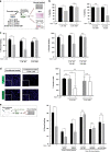
Fig. 1
Fig. 1. Secreted media from Vemurafenib-resistant melanoma cell lines induces a resistance phenotype in sensitive melanoma cells.
a Schematic representation of the in vitro assays. b Cell viability assay or c Wound healing of M3 and Sk-Mel-28 cells after being cultivated with their own conditioned media (CM) and from the corresponding resistant CM treated with Vemurafenib (1 μM, 48 h). d Representative images of Hoechst nuclear staining of the M3 cell line (x10 microscopic field) and percentage (%) of invasive cells into the matrigel after transwell assay with CM (Vemurafenib 1 μM, 48 h). e (Left) Schematic representation of the in vivo assay. (Right) Wound-healing assay of M3 cells cultivated with 10% of mice plasma from SCID mice with xenografts of A375, M238, A375R, M238R and Sk-Mel-28R melanoma cells treated with Vemurafenib (1 μM, 48 h). Graphs show mean + SEM. n > 3 independent experiments. Statistical analysis was performed using ANOVA and Bonferroni tests (*p < 0.05; **p < 0.01; ***p < 0.001; n.s., non-significant).
Fig. 2
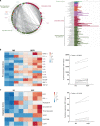
Fig. 2. Cytokine secretome profile of Vemurafenib-resistant melanoma cell lines exhibit an increase in pro-tumour cytokines.
a GO analysis based on differentially regulated cytokines on resistant melanoma cells. Each node shows an enriched GO term. Heatmap (OD mean) and cytokine expression graphic of b pro-tumour cytokine profile and c pro-angiogenic cytokine profile. Statistical analysis was performed using the Wilcoxson multiple comparison test.
Fig. 3
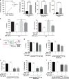
Fig. 3. M-CSF promotes the acquisition of Vemurafenib-resistance in BRAFV600E melanoma cells.
a Levels of M-CSF in conditioned media of Vem-S and Vem-R cell lines determined by ELISA assay. b Viability and wound-healing assay of sensitive melanoma cells treated with Vemurafenib (1 μM, 48 h) and/or rhM-CSF (200 ng/mL). c Schematic representation of the in vitro assays. d MTT assay or e wound-healing assay of sensitive M3 and SK-Mel-28 cells after exposure to 48 h of their corresponding resistant CM treated with Vemurafenib (1 μM) and/or M-CSF mAb (20 ng/mL). f Wound-healing assay of Sk-Mel-28 cells cultivated with 10% of mice plasma from SCID mice with Sk-Mel-28R xenografts and treated with Vemurafenib (1 μM, 48 h) and/or M-CSF mAb (20 ng/mL). Graphs show mean + SEM. n = 3 independent experiments. Statistical analysis was performed using the ANOVA test, followed by Bonferroni’s multiple comparisons test (*p < 0.05; **p < 0.01; ***p < 0.001; ns, non-significant).
Fig. 4
Fig. 4. Autophagic blockers Mibefradil/Chloroquine, Vemurafenib and M-CSF mAb-combined therapy induce apoptotic cell death and impair migration of BRAFV600E-resistant melanoma cells.
a Monitoring autophagic flux by mRFP-EGFP-LC3B assay in resistant melanoma cells after M-CSF mAb, Mibefradil or Vemurafenib treatment. b WB and quantification of LC3II protein levels from Sk-Mel-28R melanoma cell line exposed to CQ for 1 h and M-CSF mAb (20 ng/mL). c Cell viability assay, d apoptotic Annexin V assay and e wound-healing assay of Sk-Mel-28R cells treated with Vemurafenib (1 μM) and/or M-CSF mAb (20 ng/mL) and/or combined with Mibefradil (5 μM) or CQ (12.5 μM). Graphs show mean + SEM. n > 3 independent experiments. Statistical analysis was performed using the ANOVA test, followed by Bonferroni’s multiple comparisons test (*p < 0.05; **p < 0.01; ***p < 0.001; ns, non-significant).
Fig. 5
Fig. 5. Autophagic blockers Mibefradil/Chloroquine, Vemurafenib and M-CSF mAb-combined therapy reduce tumour growth and vascularisation of BRAFV600E-resistant melanoma cells
a Workflow of the in vivo experiment. (o.p. = orally; i.p. = intraperitoneal). b Tumour growth curves under treatment with Vemurafenib (50 mg/kg), CQ (30 mg/kg), M-CSF mAb (Lacnotuzumab) (4 mg/kg) or the combination therapy for 2 weeks in mice with SK-Mel-28R xenografts (n = 6). c Weight monitoring during all the following treatments. d Ki-67 Immunochemistry detection in xenografts (left) % of positive Ki-67 (right) and representative images of the Ki-67 immunostaining. e (left) % of positive immunostaining of ENG and (right) images of vascularisation represented by ENG immunostaining. Statistical analysis was performed using the ANOVA test, followed by Bonferroni’s multiple comparisons test (*p < 0.05; **p < 0.01; ***p < 0.001; ns, non-significant).
Fig. 6
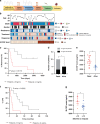
Fig. 6. M-CSF plasma levels determine melanoma OS and DFS in metastatic melanoma patients.
a Timeline of plasma collection and patient treatment. b Clinical parameters from 35 patients with BRAFV600E advanced-stage melanoma. MAP kinase-targeted therapy: BRAFi (Vemurafenib, Dabrafenib or LGX818) in monotherapy or in combination with MEKi (Dabrafenib/Trametinib, LGX818/MEK162, Vemurafenib/Cobimetinib). Response to the treatment: complete response (CR), partial responders (PR), and non-responders (stable disease (SD) and progressive disease (PD)). c Kaplan–Meier of melanoma overall survival (mOS) curve of metastatic melanoma patients (n = 17) with <5 ng/mL of M-CSF vs. >5 ng/mL. d Contingency of alive/dead patients depending on their M-CSF levels and e analysis of M-CSF plasma levels depending on the status of the patients (alive/dead). f Kaplan–Meier of melanoma disease-free survival (%DFS) of metastatic melanoma patients with <5 ng/mL vs. > 5 ng/mL of M-CSF plasma levels. g M-CSF levels depending on time to relapse after treatment (<12 vs. >12 months). Fisher exact tests were used to examine clinical variables and the ANOVA test for the statistical analysis of M-CSF plasma levels. Melanoma overall survival (mOS) and disease-free survival (DFS) curves were calculated using the Kaplan–Meier method with a log-rank test. p-values are indicated by asterisks *p < 0.05; **p < 0.01; ***p < 0.001.

References

    1. Kosmopoulou M, Giannopoulou AF, Iliou A, Benaki D, Panagiotakis A, Velentzas AD, et al. Human melanoma‐cell metabolic profiling: Identification of novel biomarkers indicating metastasis. Int J Mol Sci. 2020;21:2436. 10.3390/ijms21072436. - PMC - PubMed
    1. Mishra H, Mishra PK, Ekielski A, Jaggi M, Iqbal Z, Talegaonkar S. Melanoma treatment: from conventional to nanotechnology. J Cancer Res Clin Oncol. 2018;144:2283–302. doi: 10.1007/s00432-018-2726-1. - DOI - PMC - PubMed
    1. Trubini S, Ubiali A, Paties C, Cavanna L. Novel BRAF mutation in melanoma: a case report. Mol Clin Oncol. 2018;8:460–2. 10.3892/mco.2018.1555. Epub 2018 Jan 12. - PMC - PubMed
    1. Kakadia S, Yarlagadda N, Awad R, Kundranda M, Niu J, Naraev B, et al. Mechanisms of resistance to BRAF and MEK inhibitors and clinical update of us food and drug administration-approved targeted therapy in advanced melanoma. Onco Targets Ther. 2018;11:7095–107. doi: 10.2147/OTT.S182721. - DOI - PMC - PubMed
    1. Das A, Pushparaj C, Herreros J, Nager M, Vilella R, Portero M, et al. T-type calcium channel blockers inhibit autophagy and promote apoptosis of malignant melanoma cells. Pigment Cell Melanoma Res. 2013;26:874–85. doi: 10.1111/pcmr.12155. - DOI - PubMed

Publication types

MeSH terms