. 2022 Jun 20;13(1):3521.
doi: 10.1038/s41467-022-31166-x.

Augmented ultrasonography with implanted CMOS electronic motes

Affiliations

Augmented ultrasonography with implanted CMOS electronic motes

Yihan Zhang et al. Nat Commun. .

Abstract

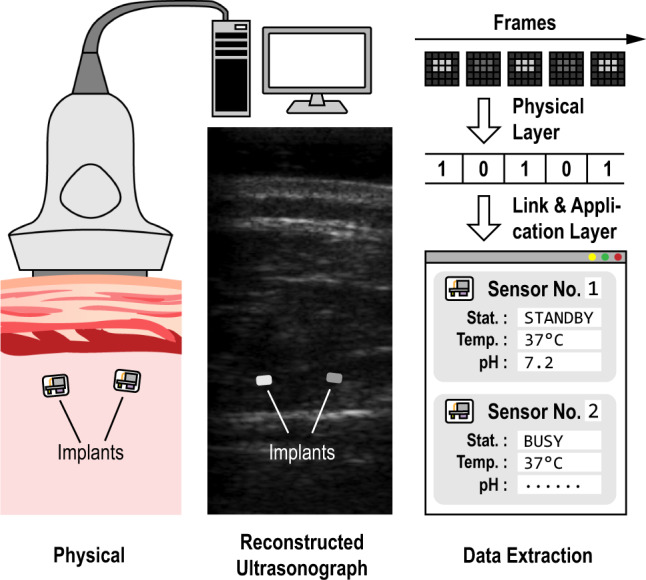
Modern clinical practice benefits significantly from imaging technologies and much effort is directed toward making this imaging more informative through the addition of contrast agents or reporters. Here, we report the design of a battery-less integrated circuit mote acting as an electronic reporter during medical ultrasound imaging. When implanted within the field-of-view of a brightness-mode (B-mode) ultrasound imager, this mote transmits information from its location through backscattered acoustic energy which is captured within the ultrasound image itself. We prototype and characterize the operation of such motes in vitro and in vivo. Performing with a static power consumption of less than 57 pW, the motes operate at duty cycles for receiving acoustic energy as low as 50 ppm. Motes within the same field-of-view during imaging have demonstrated signal-to-noise ratios of more than 19.1 dB at depths of up to 40 mm in lossy phantom. Physiological information acquired through such motes, which is beyond what is measurable with endogenous ultrasound backscatter and which is biogeographically located within an image, has the potential to provide an augmented ultrasonography.


Conflict of interest statement

The authors declare no competing interests.

Figures

Fig. 1
Fig. 1. CMOS mote implementing augmented ultrasonography.
Illustration of a typical use case; after the motes are physically implanted, they can be identified in an ultrasonograph, and data can be transmitted bidirectionally between the imager and the mote to retrieve real-time physiological information.
Fig. 2
Fig. 2. Principles of ultrasonography.
a Illustration of the pulse-echo imaging principle; b a simulated beam profile using a linear array transducer; and c measured pulse envelope at different distances from the source linear array.
Fig. 3
Fig. 3. Circuit implementation of the physical layer.
a Block diagram for the imaging system and the mote with the physical layer implementation highlighted; b top-level timing diagram of the key signals, with the pulse packet seen from the mote, assumed between 71st and 77th scanline of each frame; c the active rectifier; d the voltage regulator; e the clock recovery circuit; f the data recovery circuit; and g the uplink backscatter modulator.
Fig. 4
Fig. 4. Fabricated IC.
a Die micrograph; b an example of a processed fuse; and c a fully integrated implantable mote.
Fig. 5
Fig. 5. Characterization of the mote in a controlled ultrasound environment.
a Experimental setup and pixel intensity changes at passive structures and the active mote across frames in the reconstructed B-mode movie; b B-mode movie frames for depth characterization; and c the minimum source pressure required to properly power the mote at these depths and the detected signal-to-noise ratio in the corresponding B-mode movies.
Fig. 6
Fig. 6. Measured response from the mote in both in vitro and in vivo environments.
a Setup with two devices embedded in layers of chicken breast, with approximate mote locations overlaid; b the B-mode movie captured by the imaging system, where temporal average removal is used to reveal locations with abrupt, frame-to-frame intensity changes (plotted versus frames at the bottom); c detailed timing diagram showing instructions sent from the transducer and the motes’ replies (in frame-domain, a detected change implies a bit 0, information translated according to Supplementary Table S3/S4); d a setup with one devices implanted in the mouse’s hindlimb; e the B-mode movie and data pattern from the mote for this in vivo experiment; and f detailed timing diagram showing the interaction between the imaging system and the implanted mote.

References

    1. Wild JJ, Reid JM. Application of echo-ranging techniques to the determination of structure of biological tissues. Science. 1952;115:226–230. doi: 10.1126/science.115.2983.226. - DOI - PubMed
    1. Culjat MO, Goldenberg D, Tewari P, Singh RS. A review of tissue substitutes for ultrasound imaging. Ultrasound Med. Biol. 2010;36:861–873. doi: 10.1016/j.ultrasmedbio.2010.02.012. - DOI - PubMed
    1. Goss SA, Johnston RL, Dunn F. Comprehensive compilation of empirical ultrasonic properties of mammalian tissues. J. Acoust. Soc. Am. 1978;64:423–457. doi: 10.1121/1.382016. - DOI - PubMed
    1. Weber MJ, et al. A miniaturized single-transducer implantable pressure sensor with time-multiplexed ultrasonic data and power links. IEEE J. Solid-State Circuits. 2018;53:1089–1101. doi: 10.1109/JSSC.2017.2782086. - DOI
    1. Piech DK, et al. A wireless millimetre-scale implantable neural stimulator with ultrasonically powered bidirectional communication. Nat. Biomed. Eng. 2020;4:207–222. doi: 10.1038/s41551-020-0518-9. - DOI - PubMed

Publication types