. 2022 Dec;40(12):1855-1861.
doi: 10.1038/s41587-022-01364-5. Epub 2022 Jun 20.

Rapid biosensor development using plant hormone receptors as reprogrammable scaffolds

Affiliations

Rapid biosensor development using plant hormone receptors as reprogrammable scaffolds

Jesús Beltrán et al. Nat Biotechnol. 2022 Dec.

Abstract

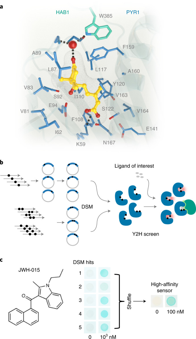
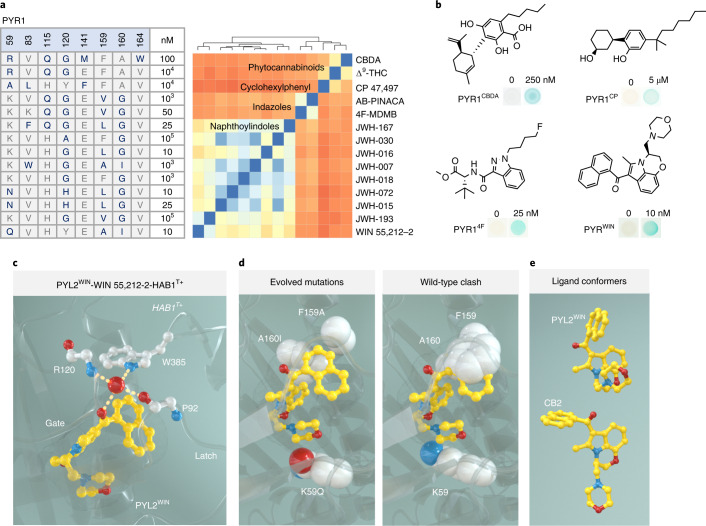
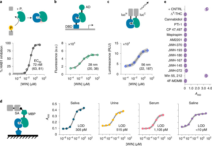
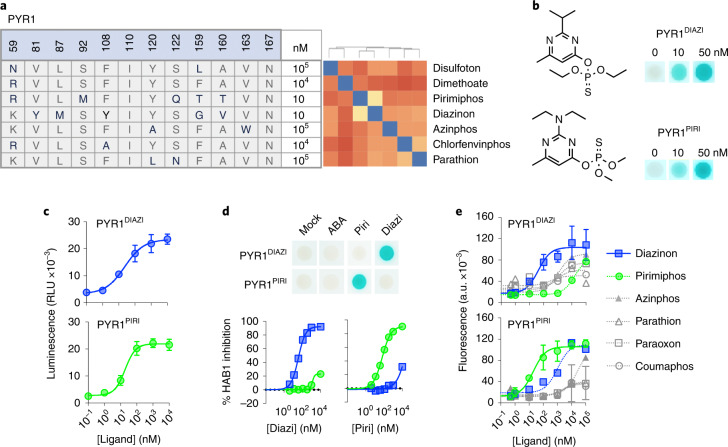
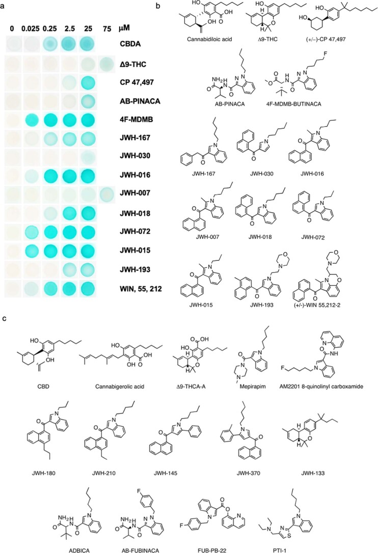
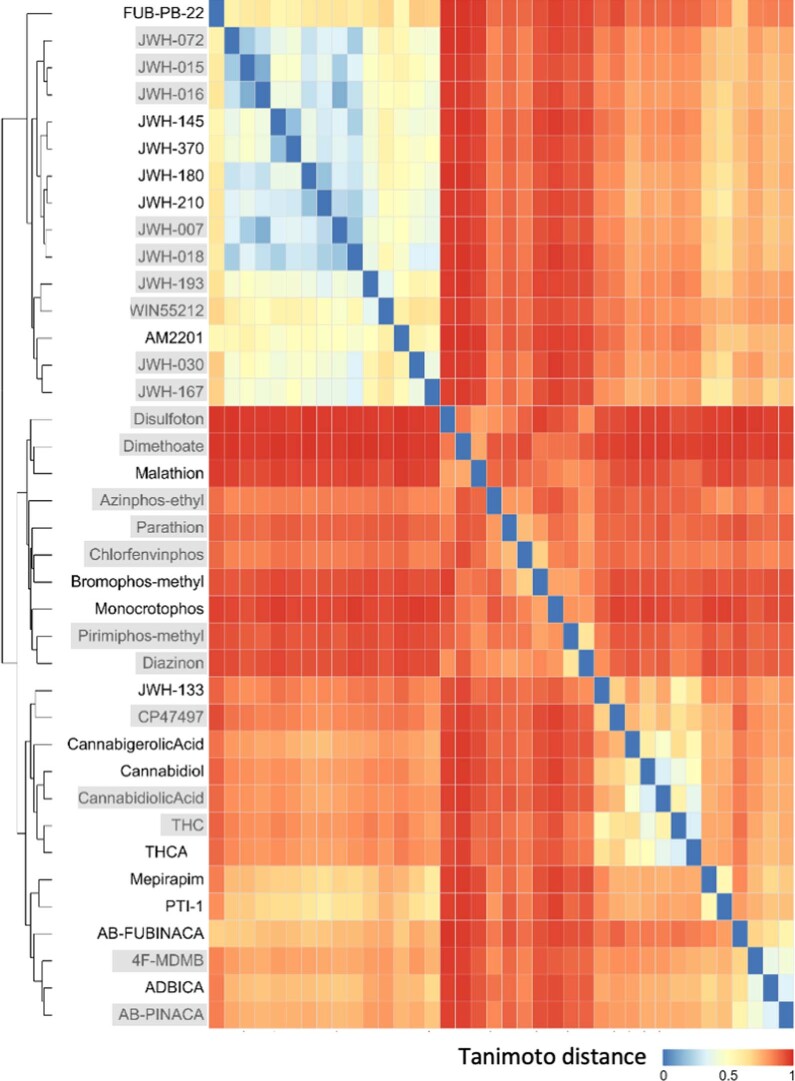
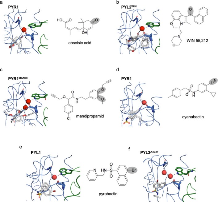
A general method to generate biosensors for user-defined molecules could provide detection tools for a wide range of biological applications. Here, we describe an approach for the rapid engineering of biosensors using PYR1 (Pyrabactin Resistance 1), a plant abscisic acid (ABA) receptor with a malleable ligand-binding pocket and a requirement for ligand-induced heterodimerization, which facilitates the construction of sense-response functions. We applied this platform to evolve 21 sensors with nanomolar to micromolar sensitivities for a range of small molecules, including structurally diverse natural and synthetic cannabinoids and several organophosphates. X-ray crystallography analysis revealed the mechanistic basis for new ligand recognition by an evolved cannabinoid receptor. We demonstrate that PYR1-derived receptors are readily ported to various ligand-responsive outputs, including enzyme-linked immunosorbent assay (ELISA)-like assays, luminescence by protein-fragment complementation and transcriptional circuits, all with picomolar to nanomolar sensitivity. PYR1 provides a scaffold for rapidly evolving new biosensors for diverse sense-response applications.


Conflict of interest statement

P.J.S., M.B., T.A.W, S.R.C., I.W. and J.B. have filed a provisional patent entitled REAGENTS AND SYSTEMS FOR GENERATING BIOSENSORS (US9738902B2; WO2011139798A2) covering the research in the present work.

Figures

Fig. 1
Fig. 1. Protein structure-guided design of high-affinity PYR1-based cannabinoid sensors.
a, The 19 side chains of residues in PYR1’s binding pocket targeted for double-site mutagenesis (DSM) are shown along with ABA (yellow) and HAB1’s W385 ‘lock’ residue and water network (3QN1). b, Sensor evolution pipeline. The PYR1 library was constructed by NM, in two subpools, one using single-mutant oligos and another using double-mutant oligo pools. The combined pools were screened for sensors using Y2H growth selections in the presence of a ligand of interest. c, Representative screen results. The DSM library was screened for mutants that respond to the synthetic cannabinoid JWH-015 yielding five hits that were subsequently optimized by two rounds of DNA shuffling to yield PYR1JWH-015, which harbors four mutations. The yeast two-hybrid (Y2H) staining data show different receptor responses to JWH-015 by β-galactosidase activity.
Fig. 2
Fig. 2. Sequence and structural basis of ligand recognition by evolved PYR1 sensors.
a, Sequence diversity of cannabinoid receptor ligand-binding pocket residues (mutant residues are shown in bold type). The minimal ligand concentrations required for Y2H signal generation are indicated at right (Supplementary Fig. 2 shows full data, including mutations outside the pocket). The heatmap shows the ligands screened clustered by their pairwise Tanimoto distance scores calculated using ChemMine; blue indicates high similarity, and orange has lower similarity. b, Representative optimized sensor Y2H β-galactosidase responses to the ligands indicated; PYR1CBDA was evolved for recognition of CBDA, PYR1CP for CP 47,497, PYR14F for 4F-MDMB and PYR1WIN for WIN 55,212-2. ce, Structural basis for cannabinoid recognition. c, WIN is colored yellow, and key ligand-contacting residues are indicated with dashes. The Trp-lock water network that stabilizes binding is shown at top. d, Relief of steric clash by the evolved receptor. e, Structural poses of WIN in PYL2-bound (top) and CB2-bound (bottom, 6PT0) structures.
Fig. 3
Fig. 3. PYR1-based sensors are portable to diverse CID-based output systems demonstrated with PYR1WIN.
a, Phosphatase inhibition. Ligand-dependent inhibition of ΔN-HAB1 phosphatase activity by recombinant receptors using a fluorogenic substrate. Inhibition expressed relative to mock controls (n = 3). b, Gene activation. Ligand-induced gene activation in S. cerevisiae using an engineered Z4-PYR1/VP64-ΔN-HAB1 genetic circuit. Whole-cell fluorescence generated from an integrated Z44-CYC1core-GFP-CYC1t reporter is shown (12 h after ligand addition; n = 3). c, Split luciferase complementation. Addition of ligand results in luminescence from NLucN-PYR1/NLucC-ΔN-HAB1 (n = 3). d, PYR1 ELISA-like immunoassays. Immobilized receptors recruit biotinylated ΔN-HAB1T+ in response to ligand, and colorimetric signal is generated by a secondary streptavidin-HRP conjugate. Assays conducted in fivefold dilutions of saliva, urine, serum and blank saline are shown, with the lower limit of detection (LOD; Methods) of each assay shown (n = 3). Data points represent the mean, and the 95% confidence interval is shown on fits in ad as gray shading and stated in square brackets along with the EC50 values. e, Receptor cross-reactivity evaluation in PYR1 ELISAs. The cannabinoids shown were assayed for signal generation at 2 µM. + CNTRL, PYR1M tested with 2 µM ABA (n = 3); RLU, relative luminescence unit. Protein parts: DBD, DNA binding domain; AD, activation domain; MBP, maltose binding protein; SA, streptavidin. Chemicals: THC, tetrahydrocannabinol; WIN, (+)-WIN 55,212-2. Source data
Fig. 4
Fig. 4. Facile development of potent, selective and portable organophosphate sensors.
a, Summary of biosensor screening results for a panel of ten organophosphates. The compounds screened are clustered by similarity (blue indicates more similar) using a distance matrix of pairwise Tanimoto similarity scores, calculated in ChemMine 19. The molecules that yielded hits are shown in bold type; the minimal ligand concentrations required for Y2H signal generation for optimized receptors (Methods) are indicated (Supplementary Fig. 12 shows additional details). b, The optimized PYR1DIAZI and PYR1PIRI are high-affinity sensors. Optimized receptors were tested for responses to nanomolar concentrations of diazinon and pirimiphos-methyl, respectively, as evidenced by Y2H assays and receptor-mediated inhibition of HAB1 phosphatase activity in vitro. PYR1DIAZI (EC50 = 36 nM [32,40]); PYR1PIRI (EC50 = 58 nM [50,67]). Wild-type PYR1 was used as a control (gray lines). c, PYR1-derived receptors are portable. PYR1DIAZI and PYR1PIRI were tested in a protein-fragment complementation system based on split luciferase reconstitution with NLucN-PYR1/NLucC-HAB1 fusions in yeast (PYR1DIAZI, EC50 = 24 nM [12,50]; PYR1PIRI, EC50 = 19 nM [undef, 29]). d,e, PYR1DIAZI and PYR1PIRI are selective for their evolved target ligands. d, Y2H (top) and in vitro phosphatase inhibition assays (bottom) were used to profile receptor responses; the receptors no longer bind the native ligand ABA. Pirimiphos-methyl and diazinon were tested 20 nM, ABA, tested at 5,000 nM in Y2H assays. e, Characterization of receptor selectivity using a Z4-PYR1/VP64-ΔN-HAB1 gene activation circuit in the presence of the activating ligands shown (Supporting File 1 shows quantitative analyses of EC50 values). In all cases, the symbol represents the mean, and the error bars show 1 s.d. and may be smaller than the symbol. All data points represent the mean of triplicate data (n = 3), and error bars represent the standard deviation. Source data
Extended Data Fig. 1
Extended Data Fig. 1. Chemical diversity of natural and synthetic cannabinoids sensed by engineered PYR1 sensors.
(a) Y2H data of sensor hits; each row shows the response of an evolved PYR1 sensor to its target ligand; the sequences of the sensors used are shown in supporting data file 1. (b) Chemical structures of compounds for which at least one PYR1-based biosensor was identified. (c) Chemical structures of compounds for which no PYR1-based biosensor was identified.
Extended Data Fig. 2
Extended Data Fig. 2. Chemicals subjected to sensor screens using PYR1 DSM library.
The 38 chemicals screened in our manuscript were compared pairwise to compute a matrix of Tanimoto distance scores (shown in the heatmap), which was used for hierarchical clustering; blue = small distance (high similarity) and red = higher distance. Sensors were obtained for the 21 compounds in the shaded boxes.
Extended Data Fig. 3
Extended Data Fig. 3. Diverse H-bond acceptors can stabilize activated PYR/PYL receptors.
Diverse H-bond acceptors can stabilize activated PYR/PYL receptors. Structural comparisons of diverse receptor-ligand complexes with the receptor shown in blue and the complexed PP2C (HAB1) is shown in green. (a) In wild-type complexes, the PYR1/ABA/HAB1 complex is stabilized by a hydrogen-bond network coordinated by a central water molecule conserved across ligand-activated receptor X-ray structures (red). The conserved water stabilizes the ternary complex by making contacts to ABA’s cyclohexenone oxygen and PYR1’s P88 main-chain carbonyl oxygen, which serve as H-bond acceptors, and HAB1’s TRP385 indole HN and PYR1’s ARG116 main-chain NH, which serve as H-donors (PDB 3QN1). (b) In the engineered PYL2WIN receptor, WIN 55,212’s naphthoylindole carbonyl oxygen interacts with the conserved water molecule to engage the TRP-lock, mimicking ABA’s carbonyl oxygen. (c) In the engineered PYR1MANDI receptor, mandipropamid’s aryl-alkyl ether oxygens stabilizes the PYR1MANDI/mandipropamid/HAB1 complex via two bound waters that interact with ether oxygens (PDB 4WVO). (d) The rationally designed agonist cyanabactin stabilizes activated PYR1 via an arylnitrile H-bond acceptor that interacts with the conserved water (PDB 5UR6). (e, f) The agonist pyrabactin (with a weak C-Br H-bond acceptor) does not make appreciable contacts with bound water either in homologous PYL1 receptor nor in the engineered PYL2A393F. (e shows PDB ID 3NMT, native PYL1, PDB ID 3NMN is shown in f).

References

    1. Glasgow AA, et al. Computational design of a modular protein sense-response system. Science. 2019;366:1024–1028. doi: 10.1126/science.aax8780. - DOI - PMC - PubMed
    1. Taylor ND, et al. Engineering an allosteric transcription factor to respond to new ligands. Nat. Methods. 2016;13:177–183. doi: 10.1038/nmeth.3696. - DOI - PMC - PubMed
    1. Urban DJ, Roth BL. DREADDs (designer receptors exclusively activated by designer drugs): chemogenetic tools with therapeutic utility. Annu. Rev. Pharmacol. Toxicol. 2015;55:399–417. doi: 10.1146/annurev-pharmtox-010814-124803. - DOI - PubMed
    1. Tinberg CE, et al. Computational design of ligand-binding proteins with high affinity and selectivity. Nature. 2013;501:212–216. doi: 10.1038/nature12443. - DOI - PMC - PubMed
    1. Polizzi NF, DeGrado WF. A defined structural unit enables de novo design of small-molecule–binding proteins. Science. 2020;369:1227–1233. doi: 10.1126/science.abb8330. - DOI - PMC - PubMed

Publication types