Review
. 2023 Jan;252(1):27-60.
doi: 10.1002/dvdy.512. Epub 2022 Jul 6.

Structure and evolution of neuronal wiring receptors and ligands

Affiliations
Review

Structure and evolution of neuronal wiring receptors and ligands

Elena Cortés et al. Dev Dyn. 2023 Jan.

Abstract

One of the fundamental properties of a neuronal circuit is the map of its connections. The cellular and developmental processes that allow for the growth of axons and dendrites, selection of synaptic targets, and formation of functional synapses use neuronal surface receptors and their interactions with other surface receptors, secreted ligands, and matrix molecules. Spatiotemporal regulation of the expression of these receptors and cues allows for specificity in the developmental pathways that wire stereotyped circuits. The families of molecules controlling axon guidance and synapse formation are generally conserved across animals, with some important exceptions, which have consequences for neuronal connectivity. Here, we summarize the distribution of such molecules across multiple taxa, with a focus on model organisms, evolutionary processes that led to the multitude of such molecules, and functional consequences for the diversification or loss of these receptors.

Keywords: axon guidance; cell adhesion molecule; cell surface receptor; gene duplication; molecular evolution; secreted ligand; synaptogenesis.


Conflict of interest statement

The authors declare no conflicts of interest.

Figures

FIGURE 1
FIGURE 1
Phylogenetic tree of metazoans and their closest relatives. The three whole genome duplication events mentioned in the text are labeled with red asterisks. Some taxa are omitted for clarity. Positions of some of the branches are uncertain; for example, the positions of Porifera, Ctenophora, Cnidaria, and Placozoa are still debated within the early metazoan branches. Only Ctenophora, Cnidaria, and Bilateria have nervous systems.
FIGURE 2
FIGURE 2
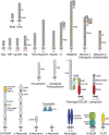
Domain architectures of axon guidance proteins. Guidance cues are listed on top and receptors are listed at the bottom. Each domain type has been drawn with a unique color and shape, and lengths of domains are approximately representative of their length in the primary sequence. Intracellular motifs mentioned in text are labeled with small purple boxes. Proteins in the top row have their N termini at the bottom, while the proteins in the bottom row have their N termini at the top. Dashed line in Slit represents proteolysis.
FIGURE 3
FIGURE 3
Domain architecture of cell adhesion molecules involved in synapse targeting, formation and function. Each domain type has been drawn with a unique color and shape (same as in Figure 2), and lengths of domains are approximately representative of their length in the primary sequence. Red stripes in Dscams and Neurexins indicate sites of high diversity created by alternative splicing. The red asterisk in the cholinesterase domain indicates that the domain is catalytically dead. Cerebellin hexamerization is mediated by disulfide bonds, formed by the N‐terminal region (indicated by green lines). The black stripe within the GAIN domain indicates the autoproteolytic site. Intracellular motifs mentioned in text are labeled with small purple boxes. All proteins have their N termini located at the top.
FIGURE 4
FIGURE 4
AlphaFold prediction of the ectodomain of an Eph from choanoflagellate Salpingoeca rosetta. (Left) Overlay of S rosetta Eph and human EphB6 ectodomains, whose sequences fail to align using BLAST with default settings. S rosetta model is produced by the ColabFold implementation of AlphaFold, while the EphB6 structure was determined using x‐ray crystallography. (Middle, Right) S rosetta Eph ectodomain (middle) has an Eph LBD domain followed by a Sushi domain, while human EphB6 ectodomain (right) has the classical architecture including an Eph LBD domain, a Sushi domain, an EGF domain and two FNIII domains (C‐terminal FNIII domain was too flexible and could not be modeled). Each domain is colored differently. Conserved disulfide linkages, a common feature of shared folds and domains, are marked by red ovals.

References

    1. Luo L. Chapter 7. Constructing the nervous system. Principles of Neurobiology. 2nd ed. New York: Garland Science; 2020. doi:10.1201/9781003053972 - DOI
    1. Shen K, Scheiffele P. Genetics and cell biology of building specific synaptic connectivity. Annu Rev Neurosci. 2010;33:473‐507. doi:10.1146/annurev.neuro.051508.135302 - DOI - PMC - PubMed
    1. de Wit J, Ghosh A. Specification of synaptic connectivity by cell surface interactions. Nat Rev Neurosci. 2016;17(1):22‐35. doi:10.1038/nrn.2015.3 - DOI - PubMed
    1. Sanes JR, Zipursky SL. Synaptic specificity, recognition molecules, and assembly of neural circuits. Cell. 2020;181(3):536‐556. doi:10.1016/j.cell.2020.04.008 - DOI - PubMed
    1. Strausfeld NJ, Hirth F. Deep homology of arthropod central complex and vertebrate basal ganglia. Science. 2013;340(6129):157‐161. doi:10.1126/science.1231828 - DOI - PubMed

Publication types

LinkOut - more resources